Departments of Urology, The First Affiliated Hospital of Fujian Medical University, Fuzhou, China.
J Cell Mol Med. 2020 Apr;24(8):4698-4706. doi: 10.1111/jcmm.15138. Epub 2020 Mar 13.
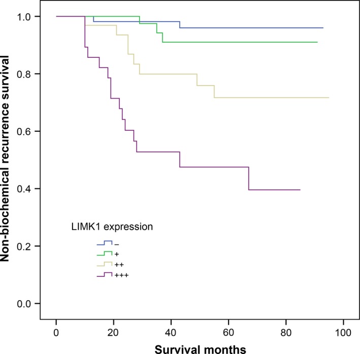
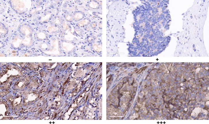
This study aimed to explore the association between LIM domain kinase 1 (LIMK1) expression in prostate cancer (PCa) tissues with advanced pathological features, lymph node metastases and biochemical recurrence. A total of 279 PCa specimens from patients who underwent radical prostatectomy and 50 benign prostatic hyperplasia (BPH) specimens were collected to construct tissue microarray, which were subjected to immunohistochemical staining for LIMK1 expression subsequently. Logistic and Cox regression analysis were used to evaluate the relationship between LIMK1 expression and clinicopathological features of patients with PCa. Immunohistochemical staining assay demonstrated that LIMK1 expression was significantly higher in PCa than BPH specimens (77.1% vs 26.0%; P < .001). LIMK1 expression was significantly higher in positive lymph node specimens than corresponding PCa specimens (P = .002; P < .001). Up-regulation of LIMK1 was associated with prostate volume, prostate-specific antigen, prostate-specific antigen density, Gleason score, T stage, lymph node metastases, extracapsular extension and seminal vesicle invasion, and positive surgical margin. Multivariate logistic regression analysis demonstrated that LIMK1 was an independent risk factor for PCa lymph node metastasis (P < .05). Multivariate Cox regression analysis revealed that the up-regulation of LIMK1 was an independent risk factor for biochemical recurrence. Kaplan-Meier analysis indicated that up-regulation LIMK1 was associated with shortened biochemical-free survival (BFS) after radical prostatectomy (P < .001). In conclusion, LIMK1 was significantly up-regulated in PCa and positive lymph node specimens and correlated with lymph node metastasis and shortened BFS of PCa. The underlying molecular mechanism of LIMK1 in PCa should be further evaluated.
本研究旨在探讨前列腺癌(PCa)组织中 LIM 结构域激酶 1(LIMK1)表达与高级别病理特征、淋巴结转移和生化复发之间的关联。共收集 279 例接受根治性前列腺切除术的 PCa 标本和 50 例良性前列腺增生(BPH)标本构建组织微阵列,随后对 LIMK1 表达进行免疫组织化学染色。采用逻辑回归和 Cox 回归分析评估 LIMK1 表达与 PCa 患者临床病理特征的关系。免疫组织化学染色检测结果显示,LIMK1 在 PCa 组织中的表达明显高于 BPH 组织(77.1%比 26.0%;P<0.001)。在有淋巴结转移的 PCa 标本中,LIMK1 的表达明显高于相应的 PCa 标本(P=0.002;P<0.001)。LIMK1 的上调与前列腺体积、前列腺特异性抗原、前列腺特异性抗原密度、Gleason 评分、T 分期、淋巴结转移、包膜外侵犯和精囊侵犯以及阳性切缘有关。多因素 logistic 回归分析表明,LIMK1 是 PCa 淋巴结转移的独立危险因素(P<0.05)。多因素 Cox 回归分析显示,LIMK1 的上调是 PCa 生化复发的独立危险因素。Kaplan-Meier 分析表明,LIMK1 的上调与根治性前列腺切除术后生化无复发生存(BFS)缩短相关(P<0.001)。总之,LIMK1 在 PCa 和阳性淋巴结标本中明显上调,与淋巴结转移和 PCa 的 BFS 缩短相关。LIMK1 在 PCa 中的潜在分子机制应进一步评估。