Elemek Eser, Agrali Omer Birkan, Kuru Bahar, Kuru Leyla
Private Practice, Istanbul, Turkey.
Department of Periodontology, Faculty of Dentistry, Marmara University, Istanbul, Turkey.
Eur J Dent. 2020 Feb;14(1):24-30. doi: 10.1055/s-0040-1701162. Epub 2020 Mar 13.
Different diagnostic criteria were used for diagnosis of peri-implant diseases. The aim of this cross-sectional study was to explore prevalence of peri-implant diseases and subclassify peri-implantitis based on different levels of radiographic and clinical findings.
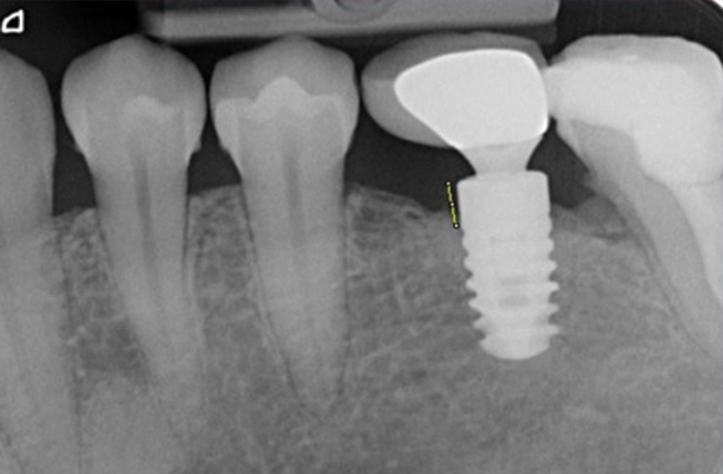
Two hundred patients having 655 dental implants were included in this study. In addition to clinical measurements, standard long-cone parallel technique was used to evaluate marginal bone level around implants. Following diagnosis of peri-implant diseases, peri-implantitis was further subclassified using a severity leveling in terms of marginal bone level and probing depth.
Mean age of 200 subjects was 52.8 ± 12.2 years and 63% were females. In total, bleeding on probing was present in 93% and suppuration in 27% of implants. On subject basis, 2.5% were diagnosed as healthy, 28% with peri-implant mucositis (PM), and 69.5% with peri-implantitis, whereas on implant basis, 3.6% were healthy, 36% presented PM, and 60.4% peri-implantitis. Furthermore, when severity leveling was applied, peri-implantitis prevalence changed markedly and ranged from 14.5 to 31.0% at the subject level and from 10.0 to 22.0% at the implant level. Subgingival restoration margins were observed in 70.6% of patients for implants with PM and in 44% patients for implants with peri-implantitis. Most of the implants with peri-implantitis were with platform match (71.5%).
Applying different thresholds to the peri-implantitis definition yielded different prevalence rates ranging from 10 to 31%. As no established diagnostic criteria are being used today, results from clinical studies may not reflect the true disease prevalence.
采用不同的诊断标准来诊断种植体周围疾病。本横断面研究的目的是探讨种植体周围疾病的患病率,并根据不同水平的影像学和临床检查结果对种植体周围炎进行亚分类。
本研究纳入了200例患者的655颗牙种植体。除临床测量外,采用标准长锥平行投照技术评估种植体周围的边缘骨水平。在诊断种植体周围疾病后,根据边缘骨水平和探诊深度的严重程度分级对种植体周围炎进行进一步亚分类。
200名受试者的平均年龄为52.8±12.2岁,63%为女性。总体而言,93%的种植体探诊出血,27%有溢脓。以受试者为基础,2.5%被诊断为健康,28%患有种植体周围黏膜炎(PM),69.5%患有种植体周围炎;而以种植体为基础,3.6%为健康,36%表现为PM,60.4%为种植体周围炎。此外,应用严重程度分级时,种植体周围炎的患病率显著变化,在受试者水平上为14.5%至31.0%,在种植体水平上为10.0%至22.0%。在患有PM的种植体患者中,70.6%观察到龈下修复边缘;在患有种植体周围炎的种植体患者中,44%观察到龈下修复边缘。大多数患有种植体周围炎的种植体为平台匹配(71.5%)。
对种植体周围炎定义应用不同阈值会产生10%至31%的不同患病率。由于目前尚未使用既定的诊断标准,临床研究结果可能无法反映真实的疾病患病率。