Wu Yufei, Tu Yunhai, Wu Chaoming, Bao Lulu, Wang Jianhua, Lu Fan, Shen Meixiao, Chen Qi
1School of Ophthalmology and Optometry, Wenzhou Medical University, 270 Xueyuan Road, Wenzhou, 325027 Zhejiang China.
2Department of Ophthalmology, Yinzhou Hospital Affiliated to Medical School of Ningbo University, Ningbo, Zhejiang China.
Eye Vis (Lond). 2020 Mar 10;7:16. doi: 10.1186/s40662-020-00180-9. eCollection 2020.
The goal was to investigate changes of the inner intra-retinal layer thicknesses and retinal capillary density (RCD) around the macula in thyroid-associated ophthalmopathy (TAO) patients with or without dysthyroid optic neuropathy (DON).
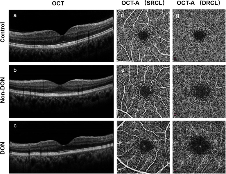
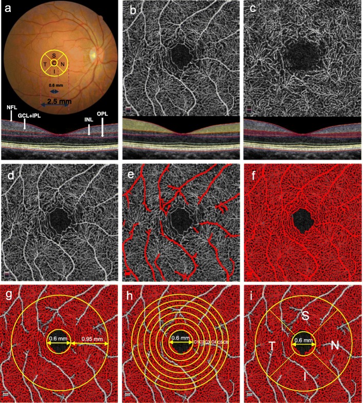
Forty-four TAO patients including 23 non-DON and 21 DON patients, and 38 healthy participants were enrolled. Spectral domain optical coherence tomography equipped with Angiovue was used to obtain three-dimensional retinal thickness maps and microvascular images of the superficial and deep retinal capillary layers (SRCL and DRCL, respectively) around the macula. Quantitative analyses were performed using a custom automated algorithm.
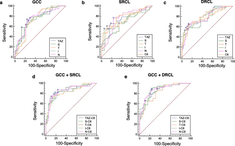
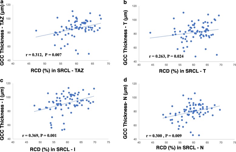
The thicknesses of the nerve fiber layer, ganglion cell layer + inner plexiform layer, and ganglion cell complex (GCC) as well as the RCDs in the SRCL and DRCL in both TAO groups were significantly decreased compared to the controls. In addition, the RCDs in DRCL of the DON group were further decreased compared to the non-DON group. GCC thickness in both TAO groups was positively correlated with the RCDs of the SRCL in the total annular zone and in the temporal, inferior, and nasal sectors. The areas under the receiver operating characteristic curves for the GCC thickness combined with the RCD were generally larger than those of each single indicator.
Thinned inner intra-retinal layers and decreased RCDs in the TAO patients without DON revealed that morphological changes might precede visual dysfunction. The composite index of the retinal structure and the microvascular density might be valuable in the diagnosing, monitoring, and intervention for early DON.
目的是研究伴有或不伴有甲状腺功能异常性视神经病变(DON)的甲状腺相关性眼病(TAO)患者黄斑周围视网膜内层厚度及视网膜毛细血管密度(RCD)的变化。
纳入44例TAO患者,其中23例无DON,21例有DON,以及38名健康参与者。使用配备Angiovue的光谱域光学相干断层扫描获取黄斑周围视网膜厚度三维图以及视网膜浅、深层毛细血管层(分别为SRCL和DRCL)的微血管图像。使用定制的自动算法进行定量分析。
与对照组相比,两个TAO组的神经纤维层、神经节细胞层+内网状层、神经节细胞复合体(GCC)厚度以及SRCL和DRCL中的RCD均显著降低。此外,与无DON组相比,有DON组的DRCL中的RCD进一步降低。两个TAO组的GCC厚度与整个环形区域以及颞侧、下方和鼻侧扇形区域的SRCL中的RCD呈正相关。GCC厚度与RCD联合的受试者工作特征曲线下面积通常大于各单一指标的曲线下面积。
无DON的TAO患者视网膜内层变薄及RCD降低表明形态学变化可能先于视功能障碍出现。视网膜结构与微血管密度的复合指标可能对早期DON的诊断、监测及干预具有重要价值。