Division of Pulmonary Medicine, Department of Medicine, Keio University School of Medicine, Tokyo 160-8582, Japan.
Division of Pulmonary Medicine, Keiyu Hospital, Yokohama, Kanagawa 220-0012, Japan.
Int J Chron Obstruct Pulmon Dis. 2020 Mar 3;15:487-499. doi: 10.2147/COPD.S230952. eCollection 2020.
Pulmonary hypertension (PH) is a major comorbidity of chronic obstructive pulmonary disease (COPD). However, the association of PH detected by echocardiography and COPD-related outcome in longitudinal follow-up has not been elucidated. In this study, we aimed to investigate the relationship between clinical characteristics of COPD patients with PH detected by echocardiography and various outcome parameters such as COPD exacerbation and health status over a three-year observation period.
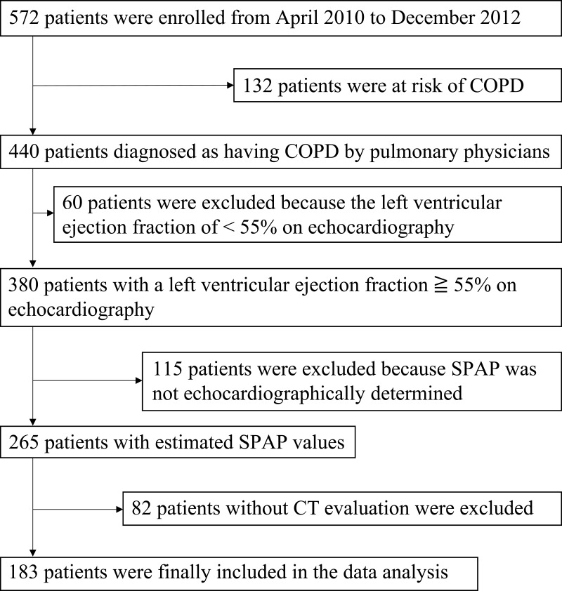
In this observational study, we analyzed patients with COPD who underwent chest computed tomography and echocardiography at baseline (n = 183).
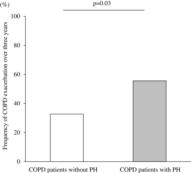
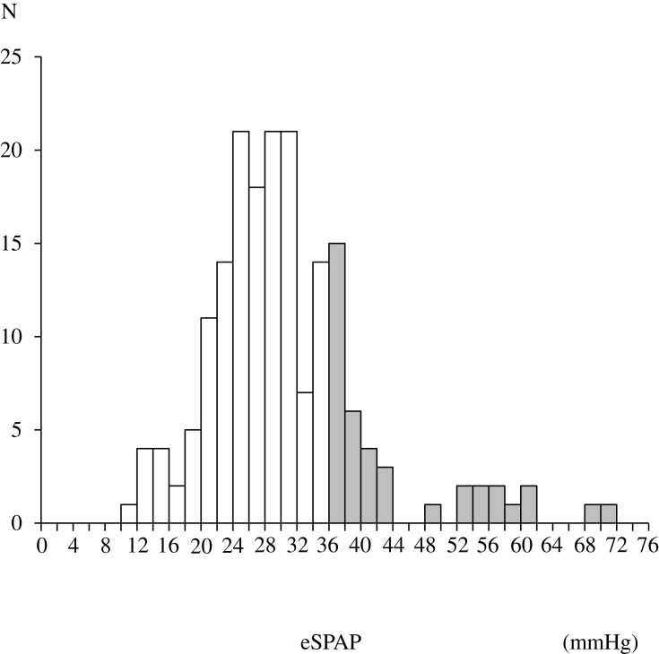
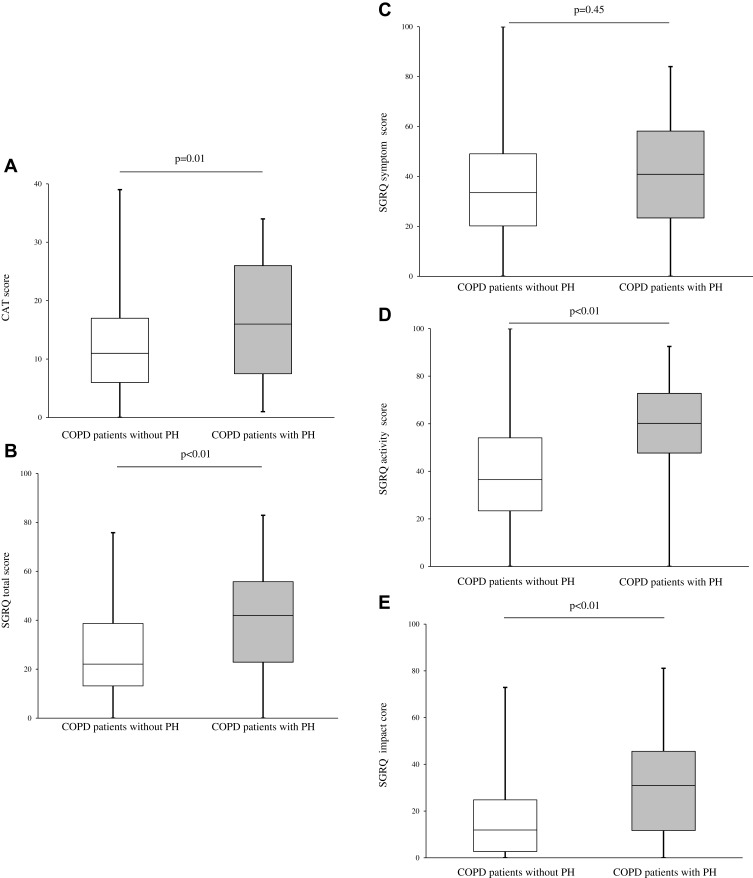
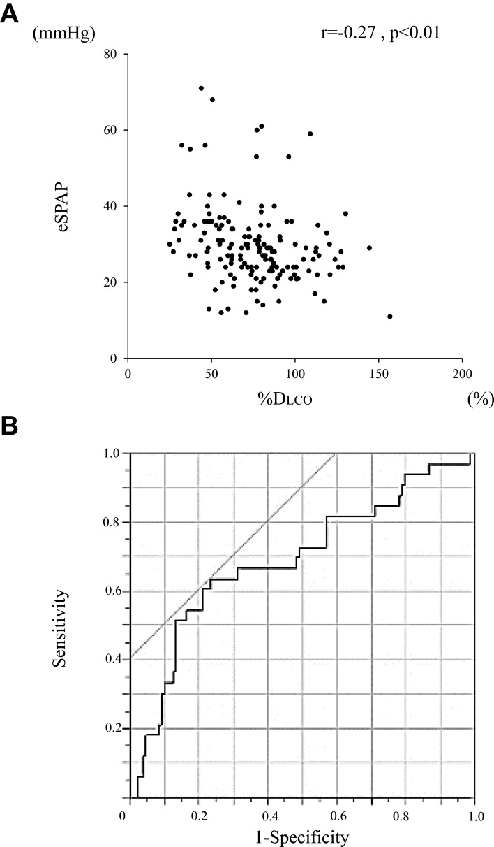
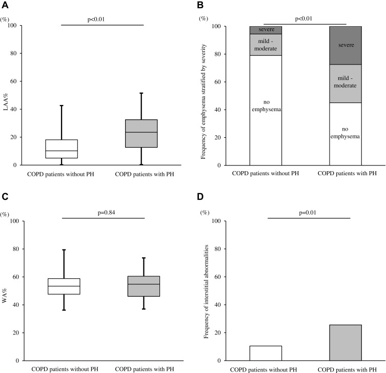
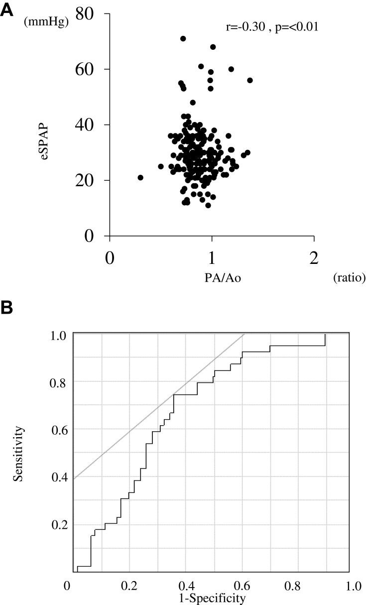
The prevalence of PH was 21.9% (40 patients). The median estimated systolic pulmonary artery pressure in patients with PH was 38.8 mmHg. COPD patients with PH were older, had a lower body mass index, scored worse in the COPD Assessment Test and St. George's Respiratory Questionnaire, and exhibited a lower diffusing capacity of the lung for carbon monoxide in comparison to patients without PH. In computed tomography images, the percentages of low-attenuation areas (LAA%) and interstitial abnormalities were higher in COPD patients with PH than in those without PH. Higher values for LAA% (LAA ≥ 30%) and interstitial abnormalities independently increased the risk of PH. The ratio of main pulmonary diameter to aortic artery diameter was significantly correlated with estimated systolic pulmonary artery pressure. In the follow-up analysis, the frequency of exacerbations in three years was significantly higher in patients with PH compared to patients without PH.
In this study, we identified the clinical characteristics of COPD patients with PH detected by echocardiography. The presence of PH assessed by echocardiography was related to future COPD exacerbations and closely related to radiographical emphysema.
肺动脉高压(PH)是慢性阻塞性肺疾病(COPD)的主要合并症。然而,超声心动图检测到的 PH 与 COPD 相关结局在纵向随访中的相关性尚未阐明。在这项研究中,我们旨在探讨超声心动图检测到的 COPD 患者的临床特征与各种结局参数之间的关系,如 COPD 加重和健康状况,观察期为三年。
在这项观察性研究中,我们分析了基线时接受胸部计算机断层扫描和超声心动图检查的 COPD 患者(n = 183)。
PH 的患病率为 21.9%(40 例)。PH 患者的中位估计收缩期肺动脉压为 38.8mmHg。与无 PH 的患者相比,PH 患者年龄更大,体重指数更低,COPD 评估测试和圣乔治呼吸问卷评分更差,一氧化碳弥散量更低。在计算机断层扫描图像中,PH 患者的低衰减区(LAA%)和间质异常百分比高于无 PH 的患者。较高的 LAA%(LAA≥30%)和间质异常独立增加 PH 的风险。主肺动脉直径与主动脉直径比值与估计的收缩期肺动脉压显著相关。在随访分析中,PH 患者在三年内的加重频率明显高于无 PH 的患者。
在这项研究中,我们确定了超声心动图检测到的 COPD 合并 PH 患者的临床特征。超声心动图评估的 PH 存在与未来 COPD 加重有关,并与影像学肺气肿密切相关。