Liu Shenhai, Jin Zhe, Zhang Yiling, Rong ShiKuo, He Wenxin, Sun Kuisheng, Wan Din, Huo Junming, Xiao Lifei, Li Xinxiao, Ding Na, Wang Feng, Sun Tao
Ningxia Key Laboratory of Cerebrocranial Disease, Incubation Base of National Key Laboratory, Ningxia Medical University, Yinchuan, China.
Department of Neurosurgery, General Hospital of Ningxia Medical University, Yinchuan, China.
Front Pharmacol. 2020 Feb 28;11:136. doi: 10.3389/fphar.2020.00136. eCollection 2020.
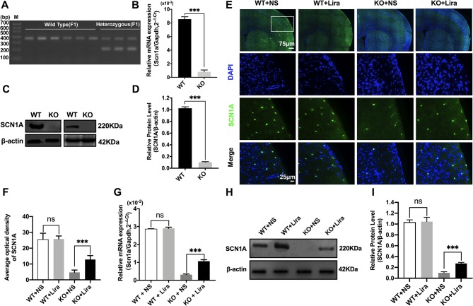
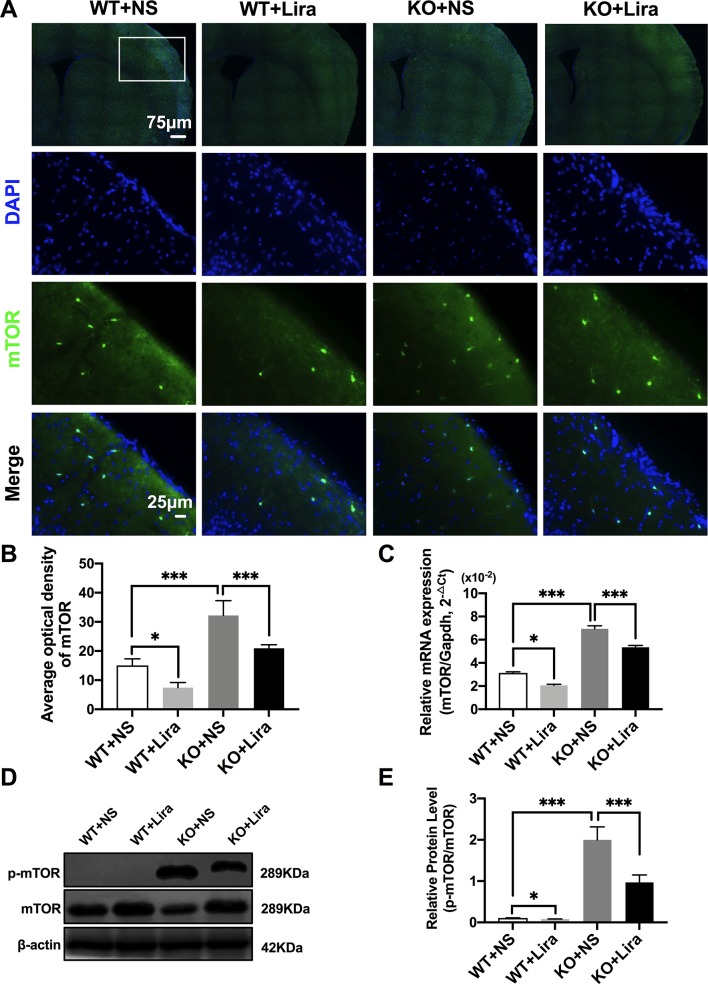
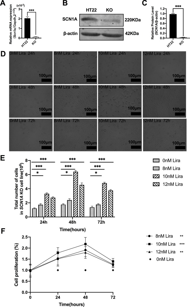
Dravet syndrome (DS) is a refractory epilepsy typically caused by heterozygous mutations of the gene, which encodes the voltage-gated sodium channel Nav1.1. Glucagon-like peptide-1 (GLP-1) analogues, effective therapeutic agents for the treatment of diabetes, have recently become attractive treatment modalities for patients with nervous system disease; however, the impact of GLP-1 analogues on DS remains unknown. This study aimed to determine the neuroprotective role of liraglutide in mouse and cell models of KO-induced epilepsy. Epileptic susceptibility, behavioral changes, and behavioral seizures were assessed using electroencephalography (EEG), IntelliCage (TSE Systems, Bad Homburg, Germany), and the open field task. Morphological changes in brain tissues were observed using hematoxylin and eosin (HE) and Nissl staining. Expression of apoptosis-related proteins and the mammalian target of rapamycin (mTOR) signaling pathway were determined using immunofluorescence and western blotting in KO-induced epileptic mice . KO model cell proliferation was evaluated using the Cell Counting Kit-8 assay, and the effect of liraglutide on cellular apoptosis levels was examined using Annexin V-FITC/PI flow cytometry. Apoptotic signal proteins and mTOR were assessed using reverse transcription - quantitative polymerase chain reaction (RT-qPCR) and western blotting. Our results showed that liraglutide significantly increased mRNA ((0.31 ± 0.04) *10 vs. (1.07 ± 0.08) * 10, = 0.0004) and protein (0.10 ± 0.02 0.27 ± 0.02, = 0.0006) expression of in KO-induced epileptic mice. In addition, liraglutide significantly alleviated electroencephalographic seizures, the severity of responses to epileptic seizures (96.53 ± 0.45 % vs. 85.98 ± 1.24 %, = 0.0003), cognitive dysfunction, and epileptic-related necrotic neurons (9.76 ± 0.91 % vs. 19.65 ± 2.64 %, = 0.0005) in KO-induced epileptic mice. Moreover, liraglutide protected against KO-induced apoptosis, which was manifested in the phosphorylation of mTOR (KO+NS: 1.99 ± 0.31 vs. KO+Lira: 0.97 ± 0.18, = 0.0004), as well as the downregulation of cleaved caspase-3 (KO+NS: 0.49 ± 0.04 vs. KO+Lira: 0.30 ± 0.01, = 0.0003) and restoration of the imbalance between BAX (KO+NS: 0.90 ± 0.02 vs. KO+Lira: 0.75 ± 0.04, = 0.0005) and BCL-2 (KO+NS: 0.46 ± 0.02 vs. KO+Lira: 0.61 ± 0.02, = 0.0006). Collectively, these results show that liraglutide reduces seizure susceptibility and cognitive dysfunction in the mouse model of Dravet syndrome, and exerts anti-apoptotic and neuroprotective effects in KO mice and cells.
德雷维特综合征(DS)是一种难治性癫痫,通常由编码电压门控钠通道Nav1.1的基因杂合突变引起。胰高血糖素样肽-1(GLP-1)类似物是治疗糖尿病的有效药物,最近已成为神经系统疾病患者有吸引力的治疗方式;然而,GLP-1类似物对DS的影响尚不清楚。本研究旨在确定利拉鲁肽在敲除(KO)诱导的癫痫小鼠和细胞模型中的神经保护作用。使用脑电图(EEG)、智能笼(TSE Systems,德国巴特洪堡)和旷场试验评估癫痫易感性、行为变化和行为性癫痫发作。使用苏木精和伊红(HE)染色及尼氏染色观察脑组织的形态学变化。在敲除诱导的癫痫小鼠中,使用免疫荧光和蛋白质印迹法测定凋亡相关蛋白的表达及雷帕霉素靶蛋白(mTOR)信号通路。使用细胞计数试剂盒-8检测法评估敲除模型细胞增殖,并使用膜联蛋白V-异硫氰酸荧光素/碘化丙啶(Annexin V-FITC/PI)流式细胞术检测利拉鲁肽对细胞凋亡水平的影响。使用逆转录-定量聚合酶链反应(RT-qPCR)和蛋白质印迹法评估凋亡信号蛋白和mTOR。我们的结果表明,利拉鲁肽显著增加敲除诱导的癫痫小鼠中某物质(原文此处未明确指出具体物质)的mRNA表达((0.31 ± 0.04)×10 vs. (1.07 ± 0.08)×10,P = 0.0004)和蛋白质表达(0.10 ± 0.02对0.27 ± 0.02,P = 0.0006)。此外,利拉鲁肽显著减轻脑电图癫痫发作、癫痫发作反应的严重程度(96.53 ± 0.45%对85.98 ± 1.24%,P = 0.0003)、认知功能障碍以及敲除诱导的癫痫小鼠中癫痫相关的坏死神经元(9.76 ± 0.91%对19.65 ± 2.64%,P = 0.0005)。此外,利拉鲁肽可防止敲除诱导的细胞凋亡,这表现为mTOR的磷酸化(敲除+生理盐水组:1.99 ± 0.31对敲除+利拉鲁肽组:0.97 ± 0.18,P = 0.0004),以及裂解的半胱天冬酶-3的下调(敲除+生理盐水组:0.49 ± 0.04对敲除+利拉鲁肽组:0.30 ± 0.01,P = 0.0003)和BAX(敲除+生理盐水组:0.90 ± 0.02对敲除+利拉鲁肽组:0.75 ± 0.04,P = 0.0005)与BCL-2(敲除+生理盐水组:0.46 ± 0.02对敲除+利拉鲁肽组:0.61 ± 0.02,P = 0.0006)之间失衡的恢复。总的来说,这些结果表明利拉鲁肽降低了德雷维特综合征小鼠模型中的癫痫易感性和认知功能障碍,并在敲除小鼠和细胞中发挥抗凋亡和神经保护作用。