Liu Qiyu, Zhou Xiaobo, Feng Wei, Pu Tao, Li Xiaoping, Li Fuyou, Kang Yu, Zhang Xiaoyan, Xu Congjian
Obstetrics and Gynecology Hospital, Fudan University, Shanghai, China.
Department of Obstetrics and Gynecology of Shanghai Medical School, Fudan University, Shanghai, China.
Front Oncol. 2020 Feb 28;10:266. doi: 10.3389/fonc.2020.00266. eCollection 2020.
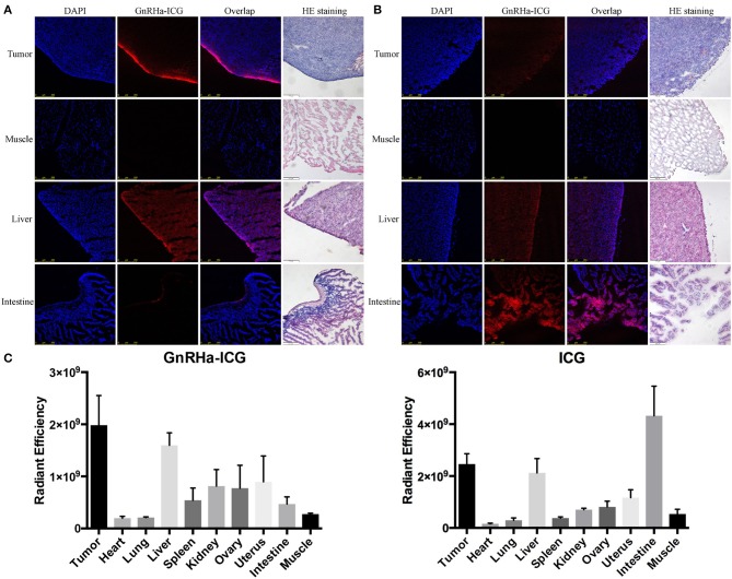
Peritoneal dissemination is common in advanced ovarian cancer. The completeness of cytoreduction is an independent prognostic factor. The intraoperative fluorescence imaging via tumor-specific near-infrared fluorophore might improve staging and surgical completeness. A promising target for ovarian cancer is the gonadotropin-releasing hormone receptor (GnRHR). This study aimed to develop a GnRHR-targeted near-infrared imaging probe for the detection of peritoneal metastases of ovarian cancer. Indocyanine green (ICG) was conjugated with GnRH antagonist peptide to develop an ovarian cancer-selective fluorescence probe GnRHa-ICG. GnRHR expression was detected in ovarian cancer tissues. The binding capacity of GnRHa-ICG and ICG was detected in both cancer cell lines and mouse models of peritoneal metastatic ovarian cancer using fluorescence microscopy, flow cytometry, and near-infrared fluorescence imaging. Tissue microarray analysis revealed the overexpression of GnRHR in ovarian cancer. GnRH-ICG exhibited the binding capacity in a panel of cancer cell lines with different expression levels of GnRHR. In ovarian cancer mouse models, GnRHa-ICG signals were detected in peritoneal tumor lesions rather than normal peritoneal and intestines tissues. ICG showed intensive fluorescence signals in intestines. The tumor-to-muscle ratio and tumor-to-intestine ratio of GnRHa-ICG was 7.41 ± 2.82 and 4.37 ± 1.66, higher than that of ICG (4.60 ± 0.50 and 0.57 ± 0.06) at 2 h post administration. The fluorescence signal of peritoneal metastases peaked in intensity at 2 h and maintained for up to 48 h. ICG also showed a weak signal in the tumor lesions due to the enhanced permeability and retention effect, but the intensity decreased quickly within 48 h. The developed GnRHR-targeted imaging agent GnRHa-ICG could specifically detected peritoneal tumor lesions from normal peritoneal and intestines tissues because of the modification of GnRHa to ICG. The plateau period of GnRHa-ICG accumulation may be feasible for clinical applications in fluorescence-guided surgery. Our GnRHR imaging concept may be effective in other hormone-related tumors with upregulated GnRHR expression.
腹膜播散在晚期卵巢癌中很常见。肿瘤细胞减灭术的彻底性是一个独立的预后因素。通过肿瘤特异性近红外荧光团进行术中荧光成像可能会改善分期和手术彻底性。卵巢癌一个有前景的靶点是促性腺激素释放激素受体(GnRHR)。本研究旨在开发一种用于检测卵巢癌腹膜转移灶的靶向GnRHR的近红外成像探针。将吲哚菁绿(ICG)与GnRH拮抗剂肽偶联,以开发一种卵巢癌选择性荧光探针GnRHa-ICG。检测卵巢癌组织中的GnRHR表达。使用荧光显微镜、流式细胞术和近红外荧光成像在癌细胞系和腹膜转移性卵巢癌小鼠模型中检测GnRHa-ICG和ICG的结合能力。组织芯片分析显示卵巢癌中GnRHR过表达。GnRH-ICG在一组具有不同GnRHR表达水平的癌细胞系中表现出结合能力。在卵巢癌小鼠模型中,在腹膜肿瘤病灶而非正常腹膜和肠道组织中检测到GnRHa-ICG信号。ICG在肠道中显示出强烈的荧光信号。给药后2小时,GnRHa-ICG的肿瘤与肌肉比值和肿瘤与肠道比值分别为7.41±2.82和4.37±1.66,高于ICG(4.60±0.50和0.57±0.06)。腹膜转移灶的荧光信号强度在2小时达到峰值,并持续长达48小时。由于增强的渗透和滞留效应,ICG在肿瘤病灶中也显示出微弱信号,但强度在48小时内迅速下降。由于对ICG进行了GnRHa修饰,所开发的靶向GnRHR的成像剂GnRHa-ICG可以特异性地从正常腹膜和肠道组织中检测出腹膜肿瘤病灶。GnRHa-ICG积累的平台期在荧光引导手术的临床应用中可能是可行的。我们的GnRHR成像概念在其他GnRHR表达上调的激素相关肿瘤中可能是有效的。