Department of Ultrasound, The Second Affiliated Hospital of Xi'an Jiaotong University, Xi'an 710004, China.
Department of Ultrasound, Northwest Women and Children Hospital, Xi'an 710061, China.
Biomed Res Int. 2020 Feb 23;2020:8701759. doi: 10.1155/2020/8701759. eCollection 2020.
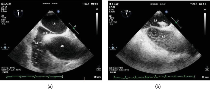
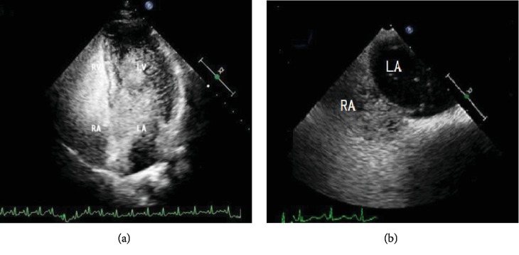
To access the distinct values of contrast transcranial Doppler (cTCD), contrast transthoracic echocardiography (cTTE), and contrast transesophageal echocardiography (cTEE) in the diagnosis of right-to-left shunt (RLS) due to patent foramen ovale (PFO) and to define the most practical strategy for the diagnosis of PFO.
102 patients with a high clinical suspicion for PFO had simultaneous cTCD, cTTE, and cTEE performed. The agitated saline mixed with blood was used to detect right-to-left shunt (RLS).
In all 102 patients, the shunt was detected at rest by cTCD in 60.78% of cases, by cTTE in 42.16%, and by cTEE in 47.06%. The positive results of all 3 techniques with Valsalva maneuver (VM) were significantly improved. cTCD showed higher pick-up rate than cTTE (98.04% vs. 89.22%; = 12.452, < 0.05) and the cTEE (98.04% vs. 96.08%; nonsignificant difference) in the diagnosis of PFO. Nevertheless, cTEE, compared with cTTE, underestimated shunting in 44% of patients. The diameter of both PFO entrance and exit was significantly greater in patients with a severe shunt compared with a mild shunt (2.8 ± 1.0 mm vs. 2.0 ± 0.7 mm, = 3.135, < 0.05) and the cTEE (98.04% vs. 96.08%; nonsignificant difference) in the diagnosis of PFO. Nevertheless, cTEE, compared with cTTE, underestimated shunting in 44% of patients. The diameter of both PFO entrance and exit was significantly greater in patients with a severe shunt compared with a mild shunt (2.8 ± 1.0 mm vs. 2.0 ± 0.7 mm, = 3.135, < 0.05) and the cTEE (98.04% vs. 96.08%; nonsignificant difference) in the diagnosis of PFO. Nevertheless, cTEE, compared with cTTE, underestimated shunting in 44% of patients. The diameter of both PFO entrance and exit was significantly greater in patients with a severe shunt compared with a mild shunt (2.8 ± 1.0 mm vs. 2.0 ± 0.7 mm, = 3.135, < 0.05) and the cTEE (98.04% vs. 96.08%; nonsignificant difference) in the diagnosis of PFO. Nevertheless, cTEE, compared with cTTE, underestimated shunting in 44% of patients. The diameter of both PFO entrance and exit was significantly greater in patients with a severe shunt compared with a mild shunt (2.8 ± 1.0 mm vs. 2.0 ± 0.7 mm.
The best method to diagnose PFO should be the combination of cTCD, cTTE, and cTEE. And cTCD should be applied as the first choice for screening RLS. Then, cTTE should be performed to quantify the severity of the shunt. Last but not least, cTEE should be performed to assess the morphologies of PFO when the closure is planned. The study provides for clinicians the most practical strategy for diagnosing PFO in the future. However, further trials with a large sample size are required to confirm this finding.
评估对比经颅多普勒超声(cTCD)、对比经胸超声心动图(cTTE)和对比经食管超声心动图(cTEE)在诊断卵圆孔未闭(PFO)所致右向左分流(RLS)中的独特价值,并确定诊断 PFO 的最实用策略。
对 102 例临床高度怀疑 PFO 的患者同时进行 cTCD、cTTE 和 cTEE 检查。采用含血生理盐水检测右向左分流(RLS)。
在所有 102 例患者中,cTCD 在静息状态下检测到分流的比例为 60.78%,cTTE 为 42.16%,cTEE 为 47.06%。所有 3 种技术在瓦氏动作(VM)下的阳性结果均显著提高。cTCD 在诊断 PFO 时的检出率高于 cTTE(98.04%比 89.22%; = 12.452,< 0.05)和 cTEE(98.04%比 96.08%;无显著差异)。然而,cTEE 比 cTTE 低估分流的比例在 44%的患者中更高。严重分流患者的 PFO 入口和出口直径明显大于轻度分流患者(2.8 ± 1.0 mm 比 2.0 ± 0.7 mm, = 3.135,< 0.05)和 cTEE(98.04%比 96.08%;无显著差异)。然而,cTEE 比 cTTE 低估分流的比例在 44%的患者中更高。严重分流患者的 PFO 入口和出口直径明显大于轻度分流患者(2.8 ± 1.0 mm 比 2.0 ± 0.7 mm, = 3.135,< 0.05)和 cTEE(98.04%比 96.08%;无显著差异)。然而,cTEE 比 cTTE 低估分流的比例在 44%的患者中更高。严重分流患者的 PFO 入口和出口直径明显大于轻度分流患者(2.8 ± 1.0 mm 比 2.0 ± 0.7 mm, = 3.135,< 0.05)和 cTEE(98.04%比 96.08%;无显著差异)。然而,cTEE 比 cTTE 低估分流的比例在 44%的患者中更高。严重分流患者的 PFO 入口和出口直径明显大于轻度分流患者(2.8 ± 1.0 mm 比 2.0 ± 0.7 mm, = 3.135,< 0.05)。
诊断 PFO 的最佳方法应是 cTCD、cTTE 和 cTEE 的联合应用。cTCD 应作为 RLS 筛查的首选方法。然后,应进行 cTTE 以量化分流的严重程度。最后但并非最不重要的是,当计划闭合时,应进行 cTEE 以评估 PFO 的形态。该研究为临床医生提供了未来诊断 PFO 的最实用策略。然而,需要进一步的大规模临床试验来证实这一发现。