Perini Alessandro, Ferrante Giada, Sivolella Stefano, Velez Joaquín Urbizo, Bengazi Franco, Botticelli Daniele
Department of Neuroscience, Division of Dentistry, University of Padua, Via Giustiniani 2, 35128, Padua, Italy.
Faculty of Dentistry, University of Medical Science, Havana, Cuba.
Int J Implant Dent. 2020 Mar 18;6(1):11. doi: 10.1186/s40729-020-0207-1.
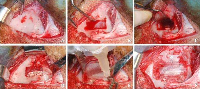
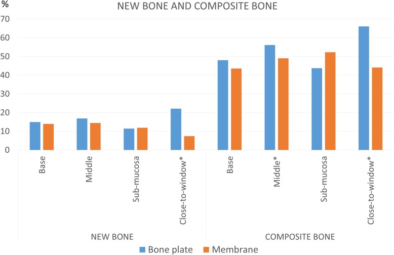
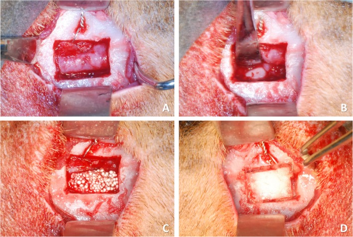
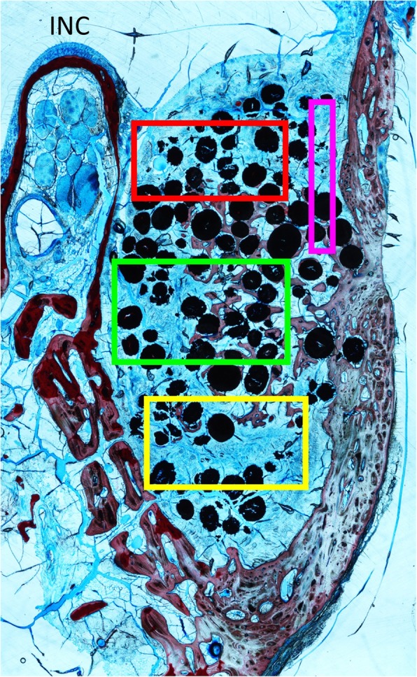
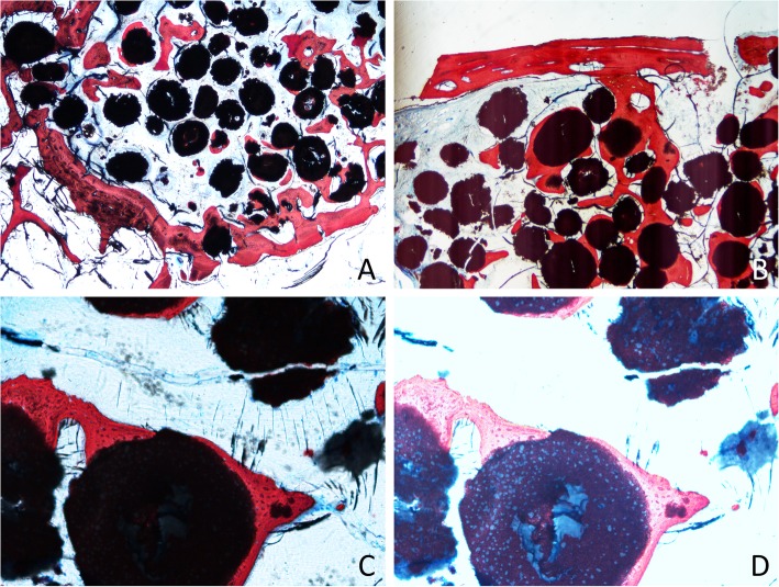
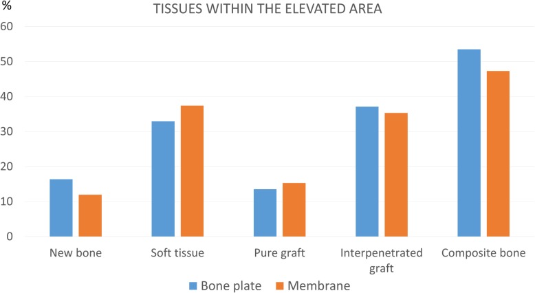
The objective of this study was to compare the healing of the augmented sinus at which the antrostomy was covered with a membrane or the repositioned bone plate.Eight sheep underwent bilateral maxillary sinus floor augmentation. The control site was covered with a resorbable membrane, while at the experimental site the bone plate was repositioned, and both were secured with cyanoacrylate. Animals were euthanised after 4 months and histomorphometric analysis was performed.A large amount of the graft appeared to be partially interpenetrated by the newly formed bone. Statistical analysis demonstrated different percentages of the new bone and bone interpenetrated to the graft between test and control site in the close-to-window area respectively 22.1 ± 12.6 vs 7.5 ± 4.5 (P = 0.028) and 66.1 ± 14.7 vs 44.2 ± 15.1 (P = 0.046). Other areas showed no difference in the bone and graft amount. More bone was found at the edges of the antrostomy in the experimental site, without statistical significance. In the centre of the antrostomy, the replaced bony window appeared bonded to the newly formed bone. No remnants and no biological response to cyanoacrylate were observed.The repositioning of the bony window after sinus floor elevation in sheep led to a larger amount of newly formed bone in the close-to-window zone of the grafted area. The bony window appeared partially bonded to the new bone. Newly formed bone was found interpenetrating the graft granules.
本研究的目的是比较用膜覆盖上颌窦开窗处或重新定位骨板时上颌窦增高后的愈合情况。八只绵羊接受了双侧上颌窦底增高术。对照部位用可吸收膜覆盖,而在实验部位重新定位骨板,两者均用氰基丙烯酸酯固定。4个月后对动物实施安乐死并进行组织形态计量学分析。大量移植物似乎被新形成的骨部分贯穿。统计分析表明,在靠近窗口区域,试验组和对照组新骨和贯穿移植物的骨的百分比不同,分别为22.1±12.6与7.5±4.5(P = 0.028)以及66.1±14.7与44.2±15.1(P = 0.046)。其他区域在骨量和移植物量方面无差异。在实验部位上颌窦开窗边缘发现更多骨,但无统计学意义。在上颌窦开窗中心,置换的骨窗似乎与新形成的骨相连。未观察到氰基丙烯酸酯的残留及生物学反应。绵羊上颌窦底提升后骨窗的重新定位导致移植区域靠近窗口区域新形成的骨量增加。骨窗似乎部分与新骨相连。发现新形成的骨贯穿移植物颗粒。