Tomita Mayu, Suzuki Motofumi, Kono Yusuke, Nakajima Kohei, Matsuda Takuma, Kuge Yuji, Ogawa Mikako
Laboratory of Bioanalysis and Molecular Imaging, Graduate School of Pharmaceutical Sciences, Hokkaido University, Sapporo, Hokkaido, 060-0812, Japan.
Central Institute of Isotope Science, Hokkaido University, Sapporo, Hokkaido, 060-0815, Japan.
EJNMMI Res. 2020 Mar 19;10(1):24. doi: 10.1186/s13550-020-0608-4.
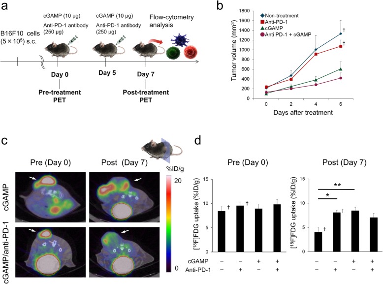
Anti-programmed cell death 1 (PD-1) antibody is an immune checkpoint inhibitor, and anti-PD-1 therapy improves the anti-tumor functions of T cells and affects tumor microenvironment. We previously reported that anti-PD-1 treatment affected tumor glycolysis by using 2-deoxy-2-[F]fluoro-D-glucose ([F]FDG) positron emission tomography (PET). That study showed that anti-PD-1 therapy in a mouse B16F10 melanoma model increased glucose metabolism in cancer cells at the point where anti-PD-1 therapy did not cause a significant inhibition of tumor growth. However, the B16F10 melanoma model is poorly immunogenic, so it is not clear how anti-PD-1 treatment affects glucose metabolism in highly immunogenic cancer models. In this study, we used a cyclic dinucleotide GMP-AMP (cGAMP)-injected B16F10 melanoma model to investigate the effect of anti-PD-1 therapy on [F]FDG uptake in a highly immune activated tumor in mice.
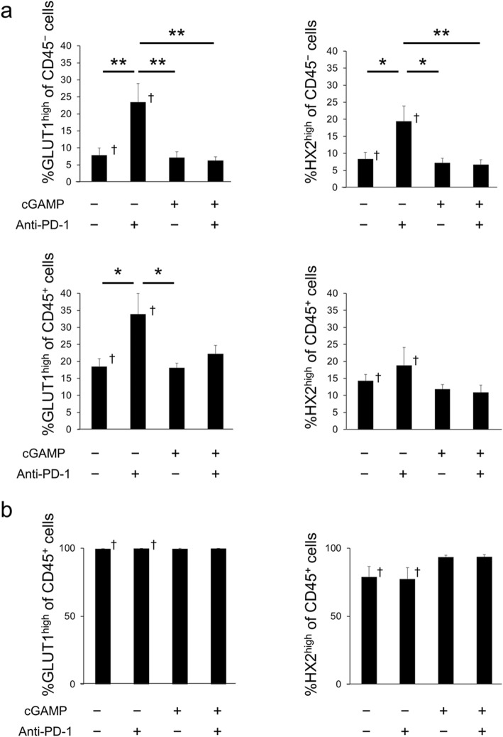
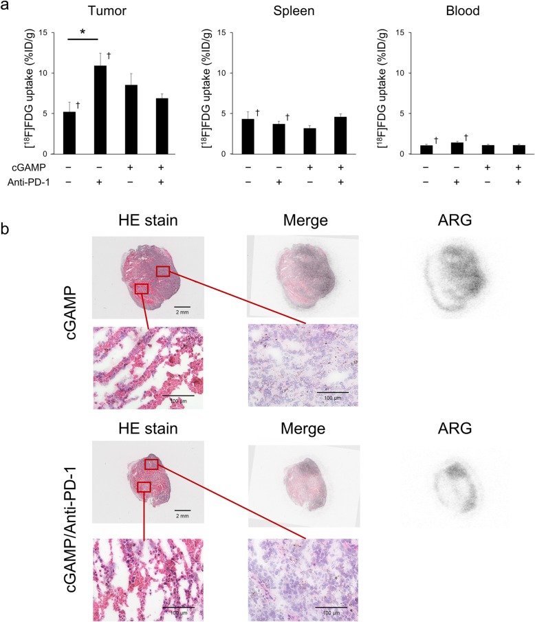
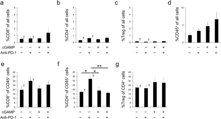
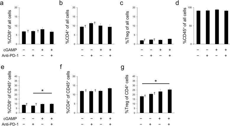
To compare the cGAMP-injected B16F10 model with the B16F10 model, experiments were performed as described in our previous manuscript. [F]FDG-PET was measured before treatment and 7 days after the start of treatment. In this study, [F]FDG uptake in tumors in the cGAMP/anti-PD-1 combination group was lower than that in the anti-PD-1 treatment group tumors on day 7, as shown by PET and ex vivo validation. Flow-cytometry was performed to assess immune cell populations and glucose metabolism. Anti-PD-1 and/or cGAMP treatment increased the infiltration level of immune cells into tumors. The cGAMP/anti-PD-1 combination group had significantly lower levels of GLUT1 cells/hexokinase II cells in CD45 cancer cells compared with tumors in the anti-PD-1 treated group. These results suggested that if immune responses in tumors are higher than a certain level, glucose uptake in cancer cells is reduced depending on that level. Such a change of glucose uptake might be caused by the difference in infiltration or activation level of immune cells between the anti-PD-1 treated group and the cGAMP/anti-PD-1 combination group.
[F]FDG uptake in cancer cells after anti-PD-1 treatment might be affected by the tumor immune microenvironment including immune cell infiltration, composition, and activation status.
抗程序性细胞死亡蛋白1(PD-1)抗体是一种免疫检查点抑制剂,抗PD-1治疗可改善T细胞的抗肿瘤功能并影响肿瘤微环境。我们之前报道过,抗PD-1治疗通过使用2-脱氧-2-[F]氟-D-葡萄糖([F]FDG)正电子发射断层扫描(PET)影响肿瘤糖酵解。该研究表明,在小鼠B16F10黑色素瘤模型中,抗PD-1治疗在未显著抑制肿瘤生长的情况下增加了癌细胞中的葡萄糖代谢。然而,B16F10黑色素瘤模型的免疫原性较差,因此尚不清楚抗PD-1治疗如何影响高免疫原性癌症模型中的葡萄糖代谢。在本研究中,我们使用环二核苷酸GMP-AMP(cGAMP)注射的B16F10黑色素瘤模型来研究抗PD-1治疗对小鼠高度免疫激活肿瘤中[F]FDG摄取的影响。
为了将cGAMP注射的B16F10模型与B16F10模型进行比较,按照我们之前论文中所述进行实验。在治疗前和治疗开始后7天测量[F]FDG-PET。在本研究中,如PET和体外验证所示,在第7天,cGAMP/抗PD-1联合治疗组肿瘤中的[F]FDG摄取低于抗PD-1治疗组肿瘤中的摄取。进行流式细胞术以评估免疫细胞群体和葡萄糖代谢。抗PD-1和/或cGAMP治疗增加了免疫细胞向肿瘤中的浸润水平。与抗PD-1治疗组的肿瘤相比,cGAMP/抗PD-1联合治疗组CD45癌细胞中GLUT1细胞/己糖激酶II细胞的水平显著降低。这些结果表明,如果肿瘤中的免疫反应高于一定水平,癌细胞中的葡萄糖摄取会根据该水平而降低。这种葡萄糖摄取的变化可能是由抗PD-1治疗组和cGAMP/抗PD-1联合治疗组之间免疫细胞浸润或激活水平的差异引起的。
抗PD-1治疗后癌细胞中的[F]FDG摄取可能受肿瘤免疫微环境影响,包括免疫细胞浸润、组成和激活状态。