Department of Anthropology, University of California San Diego, La Jolla, USA.
Institute for Neural Computation, University of California San Diego, La Jolla, USA.
Brain Struct Funct. 2020 Apr;225(3):1019-1032. doi: 10.1007/s00429-020-02055-0. Epub 2020 Mar 18.
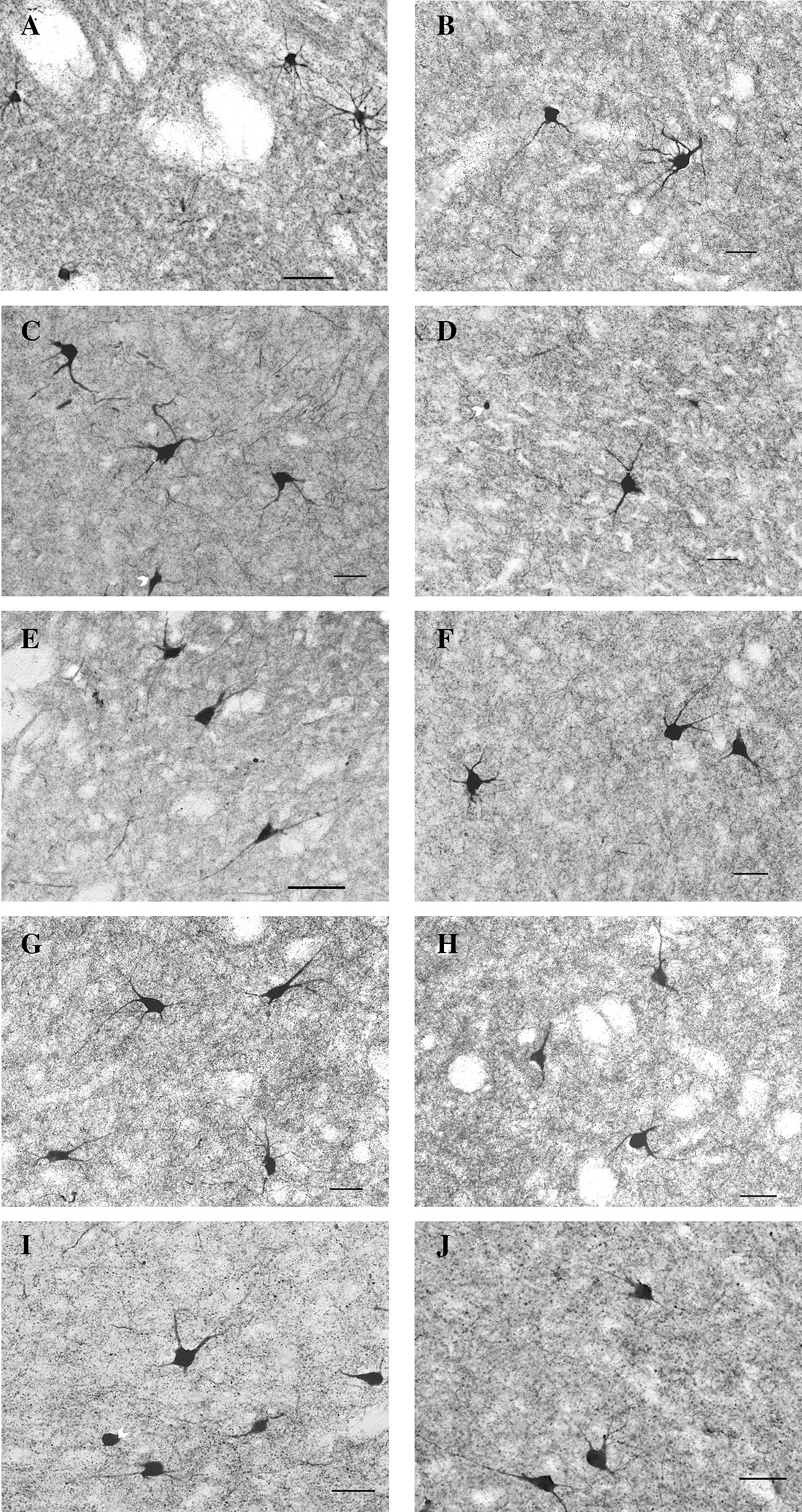
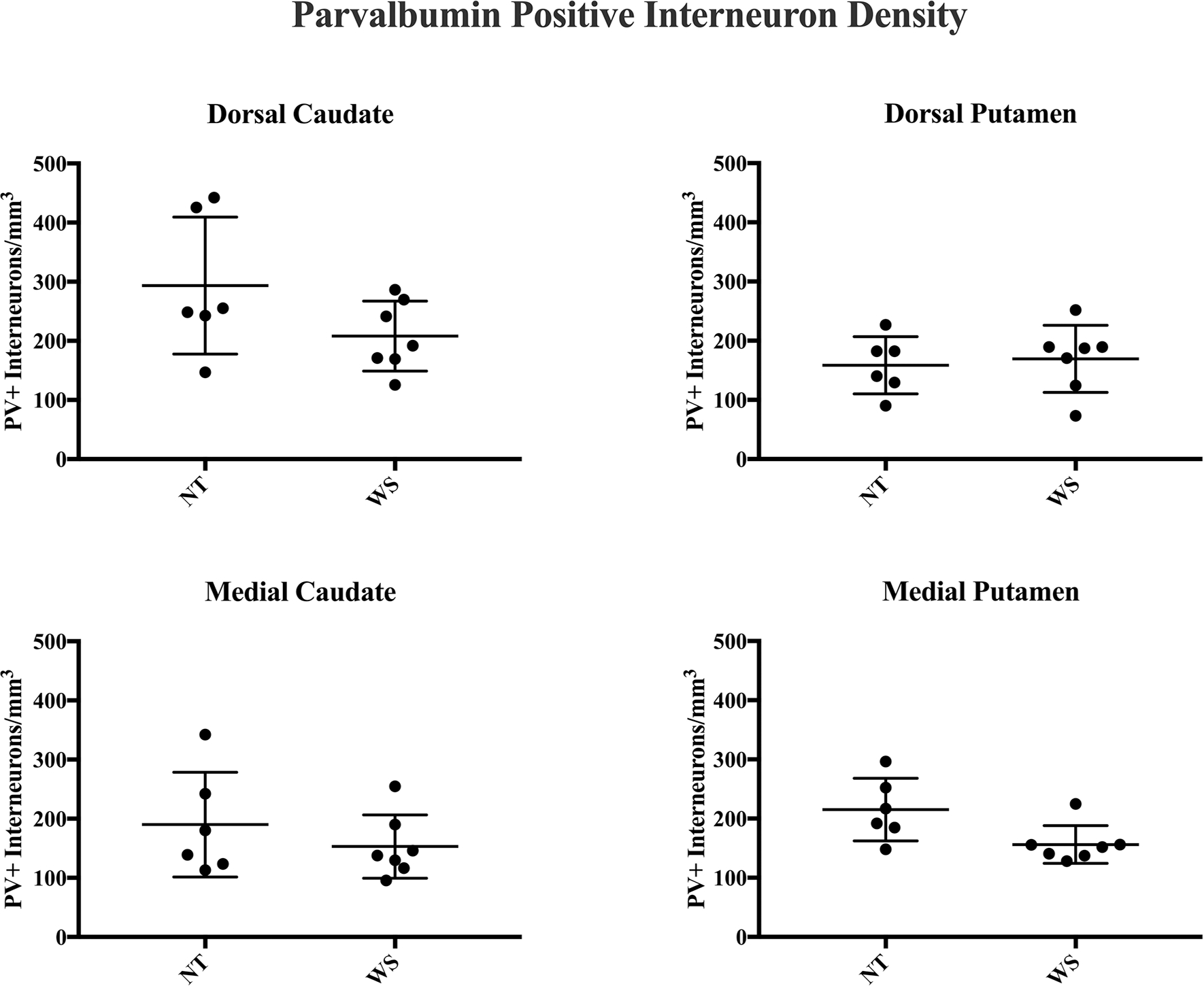
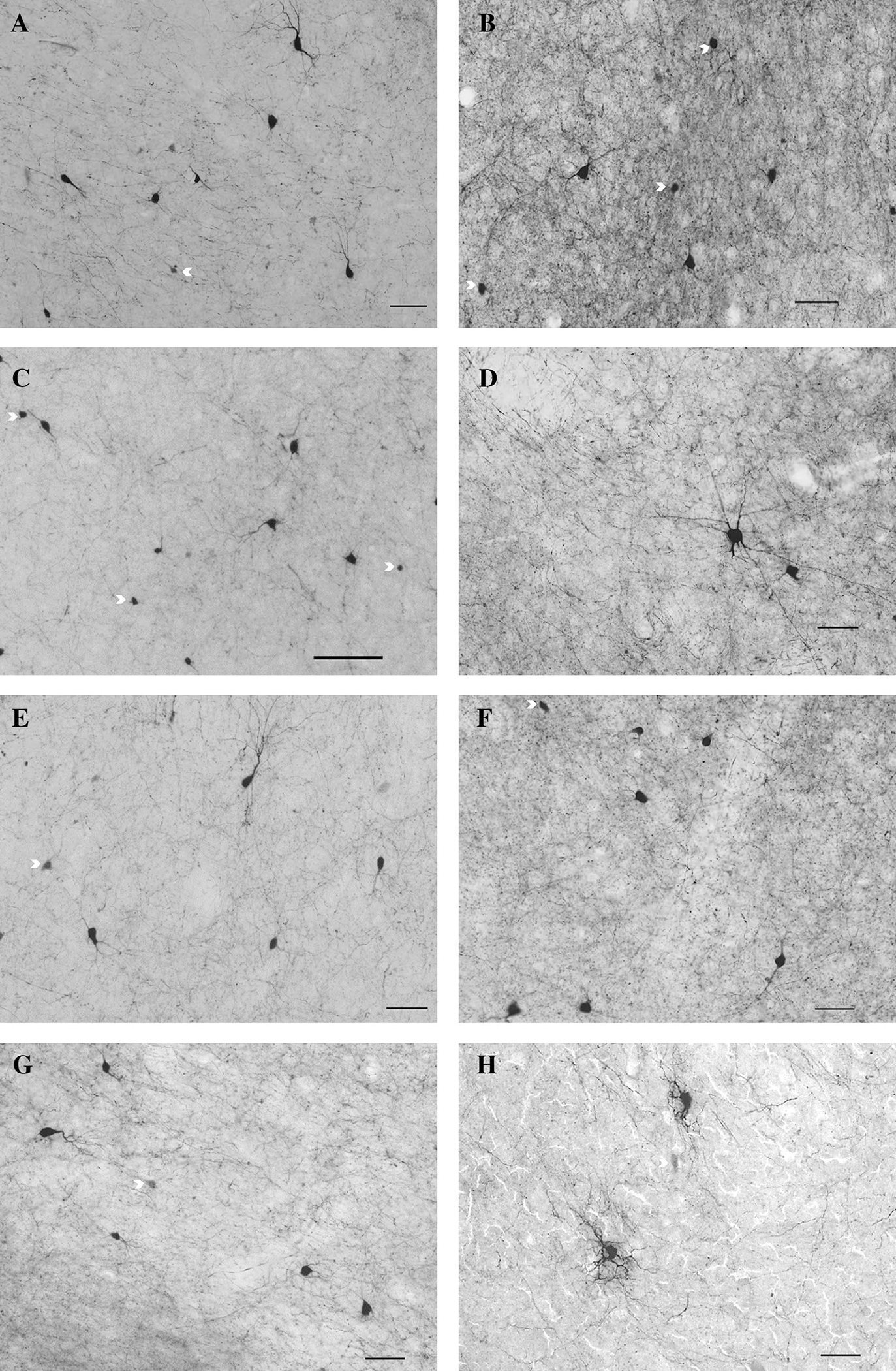
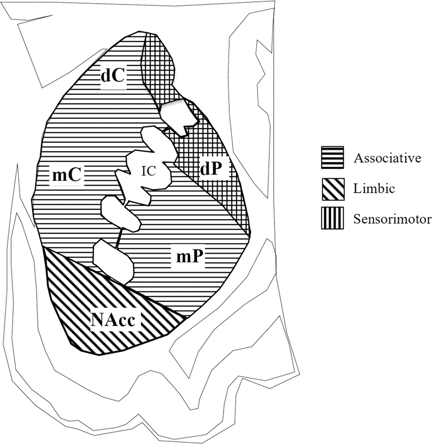
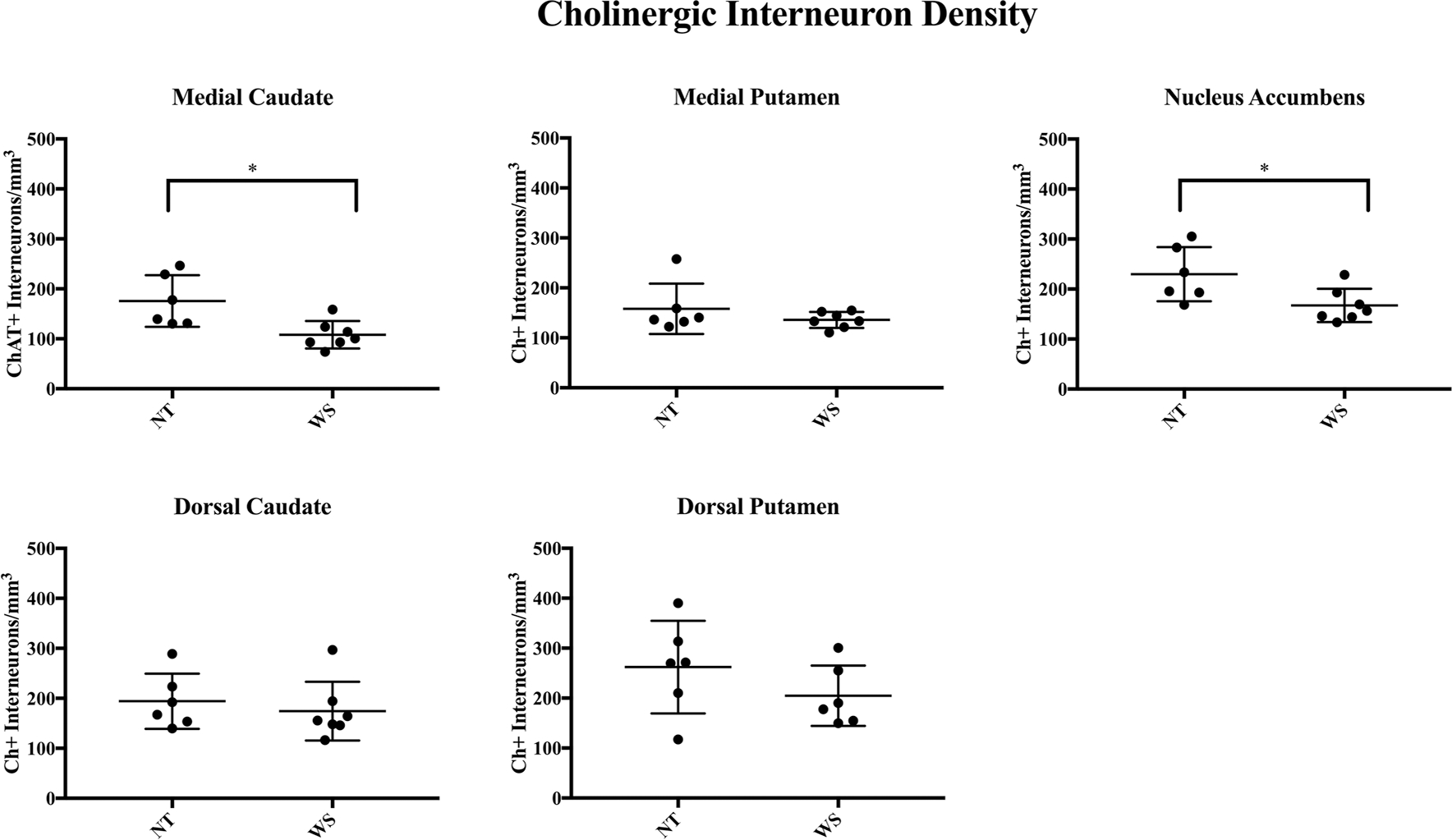
Williams syndrome (WS) is a rare neurodevelopmental disorder caused by the hemideletion of approximately 25-28 genes at 7q11.23. Its unusual social and cognitive phenotype is most strikingly characterized by the disinhibition of social behavior, in addition to reduced global IQ, with a relative sparing of language ability. Hypersociality and increased social approach behavior in WS may represent a unique inability to inhibit responses to specific social stimuli, which is likely associated with abnormalities of frontostriatal circuitry. The striatum is characterized by a diversity of interneuron subtypes, including inhibitory parvalbumin-positive interneurons (PV+) and excitatory cholinergic interneurons (Ch+). Animal model research has identified an important role for these specialized cells in regulating social approach behavior. Previous research in humans identified a depletion of interneuron subtypes associated with neuropsychiatric disorders. Here, we examined the density of PV+ and Ch+ interneurons in the striatum of 13 WS and neurotypical (NT) subjects. We found a significant reduction in the density of Ch+ interneurons in the medial caudate nucleus and nucleus accumbens, important regions receiving cortical afferents from the orbitofrontal and ventromedial prefrontal cortex, and circuitry involved in language and reward systems. No significant difference in the distribution of PV+ interneurons was found. The pattern of decreased Ch+ interneuron densities in WS differs from patterns of interneuron depletion found in other disorders.
威廉姆斯综合征(WS)是一种罕见的神经发育障碍,由 7q11.23 处约 25-28 个基因的半缺失引起。其不寻常的社会和认知表型最显著的特征是社交行为不受抑制,此外,整体智商降低,语言能力相对保留。WS 中的过度社交和增加的社交接近行为可能代表了一种无法抑制对特定社交刺激的反应的独特能力,这可能与额纹状神经回路的异常有关。纹状体的特征是具有多种中间神经元亚型,包括抑制性的 Parvalbumin 阳性中间神经元(PV+)和兴奋性的胆碱能中间神经元(Ch+)。动物模型研究已经确定了这些特化细胞在调节社交接近行为中的重要作用。以前的人类研究已经确定了与神经精神障碍相关的中间神经元亚型的耗竭。在这里,我们检查了 13 名 WS 和神经典型(NT)受试者纹状体中 PV+和 Ch+中间神经元的密度。我们发现内侧尾状核和伏隔核 Ch+中间神经元的密度显著降低,这些区域是来自眶额和腹内侧前额皮质的皮质传入的重要接收区域,并且与语言和奖励系统的回路有关。PV+中间神经元的分布没有发现显著差异。WS 中 Ch+中间神经元密度降低的模式与其他疾病中发现的中间神经元耗竭模式不同。