Hrvoj-Mihic Branka, Hanson Kari L, Lew Caroline H, Stefanacci Lisa, Jacobs Bob, Bellugi Ursula, Semendeferi Katerina
Department of Anthropology, University of California, San DiegoSan Diego, La Jolla, CA, United States.
Neuroscience Program, Colorado CollegeColorado Springs, CO, United States.
Front Neurosci. 2017 Aug 11;11:419. doi: 10.3389/fnins.2017.00419. eCollection 2017.
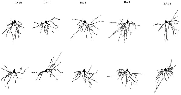
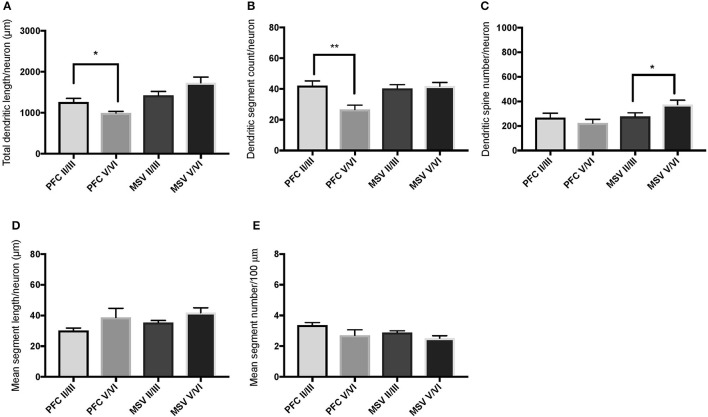
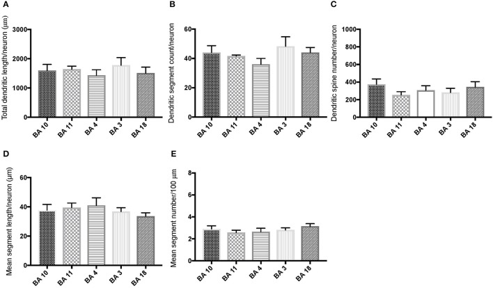
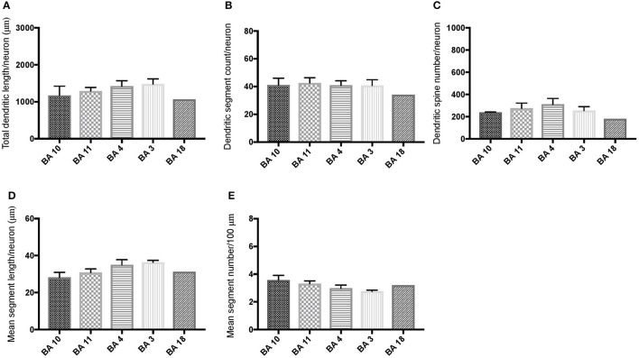
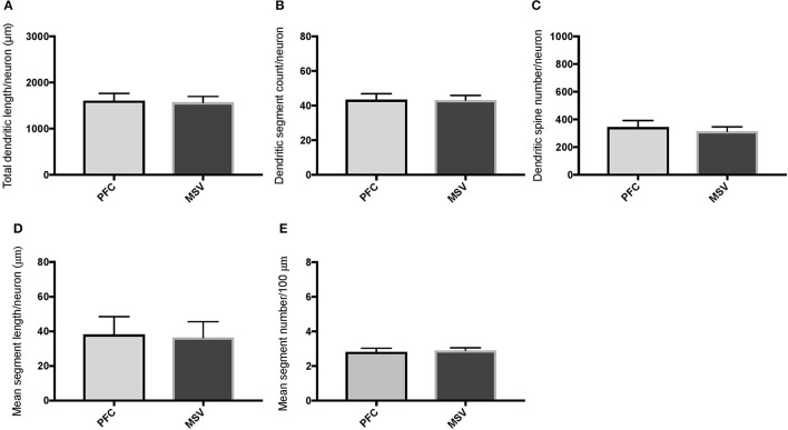
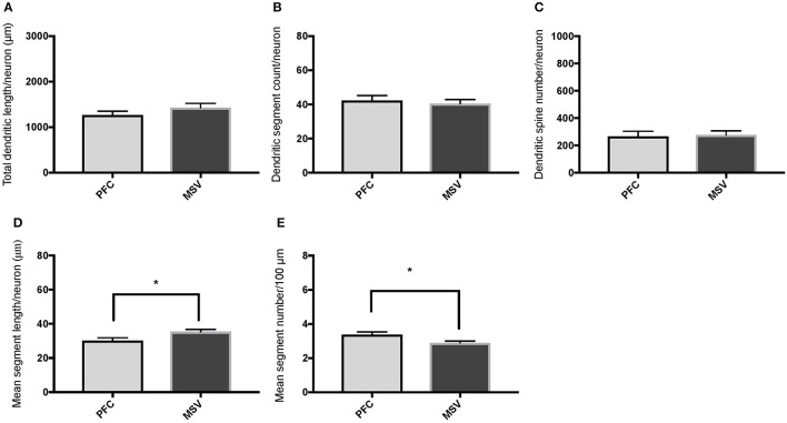
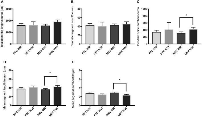
Williams syndrome (WS) is a unique neurodevelopmental disorder with a specific behavioral and cognitive profile, which includes hyperaffiliative behavior, poor social judgment, and lack of social inhibition. Here we examined the morphology of basal dendrites on pyramidal neurons in the cortex of two rare adult subjects with WS. Specifically, we examined two areas in the prefrontal cortex (PFC)-the frontal pole (Brodmann area 10) and the orbitofrontal cortex (Brodmann area 11)-and three areas in the motor, sensory, and visual cortex (BA 4, BA 3-1-2, BA 18). The findings suggest that the morphology of basal dendrites on the pyramidal neurons is altered in the cortex of WS, with differences that were layer-specific, more prominent in PFC areas, and displayed an overall pattern of dendritic organization that differentiates WS from other disorders. In particular, and unlike what was expected based on typically developing brains, basal dendrites in the two PFC areas did not display longer and more branched dendrites compared to motor, sensory and visual areas. Moreover, dendritic branching, dendritic length, and the number of dendritic spines differed little within PFC and between the central executive region (BA 10) and BA 11 that is part of the orbitofrontal region involved into emotional processing. In contrast, the relationship between the degree of neuronal branching in supra- versus infra-granular layers was spared in WS. Although this study utilized tissue held in formalin for a prolonged period of time and the number of neurons available for analysis was limited, our findings indicate that WS cortex, similar to that in other neurodevelopmental disorders such as Down syndrome, Rett syndrome, Fragile X, and idiopathic autism, has altered morphology of basal dendrites on pyramidal neurons, which appears more prominent in selected areas of the PFC. Results were examined from developmental perspectives and discussed in the context of other neurodevelopmental disorders. We have proposed hypotheses for further investigations of morphological changes on basal dendrites in WS, a syndrome of particular interest given its unique social and cognitive phenotype.
威廉姆斯综合征(WS)是一种独特的神经发育障碍,具有特定的行为和认知特征,包括过度友善行为、社会判断力差和缺乏社会抑制力。在此,我们研究了两名患有WS的成年罕见病例大脑皮层中锥体神经元基底树突的形态。具体而言,我们检查了前额叶皮层(PFC)中的两个区域——额极(布罗德曼第10区)和眶额皮层(布罗德曼第11区),以及运动、感觉和视觉皮层中的三个区域(BA4、BA3-1-2、BA18)。研究结果表明,WS患者大脑皮层中锥体神经元基底树突的形态发生了改变,这些差异具有层特异性,在前额叶皮层区域更为显著,并且呈现出一种将WS与其他疾病区分开来的整体树突组织模式。特别是,与基于正常发育大脑的预期不同,两个前额叶皮层区域的基底树突与运动、感觉和视觉区域相比,并未表现出更长且分支更多的树突。此外,在前额叶皮层内以及参与情绪处理的眶额区域的中央执行区域(BA10)和BA11之间,树突分支、树突长度和树突棘数量的差异很小。相比之下,WS患者颗粒上层和颗粒下层的神经元分支程度之间的关系未受影响。尽管本研究使用的是长期保存在福尔马林中的组织,且可供分析的神经元数量有限,但我们的研究结果表明,WS患者的大脑皮层与唐氏综合征、雷特综合征、脆性X综合征和特发性自闭症等其他神经发育障碍一样,锥体神经元基底树突的形态发生了改变,这在前额叶皮层的特定区域更为明显。我们从发育角度对结果进行了研究,并在其他神经发育障碍的背景下进行了讨论。鉴于WS独特的社会和认知表型,我们提出了一些假设,以便进一步研究WS患者基底树突的形态变化。