Zhu Wei, Chu Chengyan, Kuddannaya Shreyas, Yuan Yue, Walczak Piotr, Singh Anirudha, Song Xiaolei, Bulte Jeff W M
Russell H. Morgan Department of Radiology and Radiological Science, Division of MR Research, the Johns Hopkins University School of Medicine, Baltimore, MD, 21205, USA.
Cellular Imaging Section, Institute for Cell Engineering, the Johns Hopkins University School of Medicine, Baltimore, MD, 21205, USA.
Adv Funct Mater. 2019 Sep 5;29(36). doi: 10.1002/adfm.201903753. Epub 2019 Jul 8.
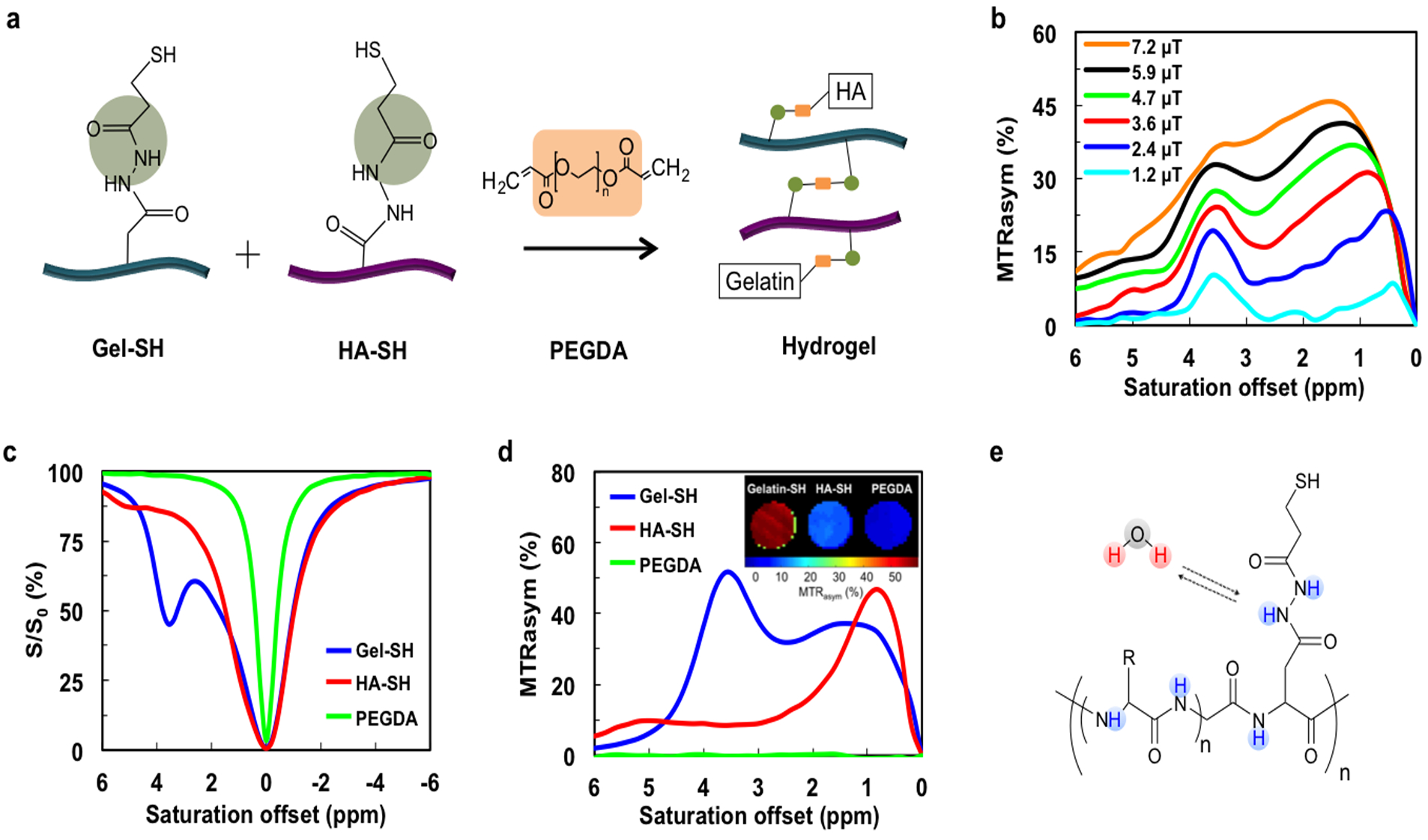
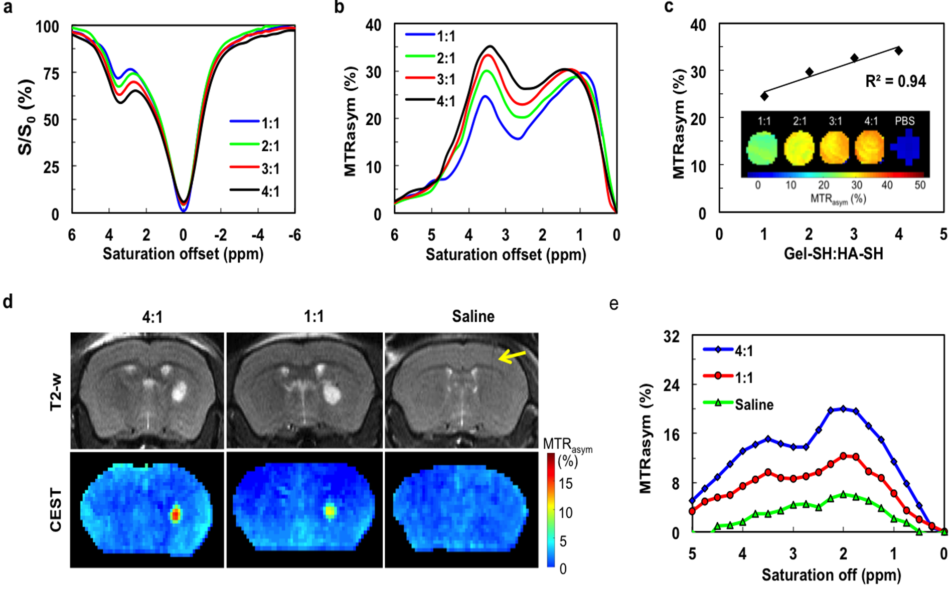
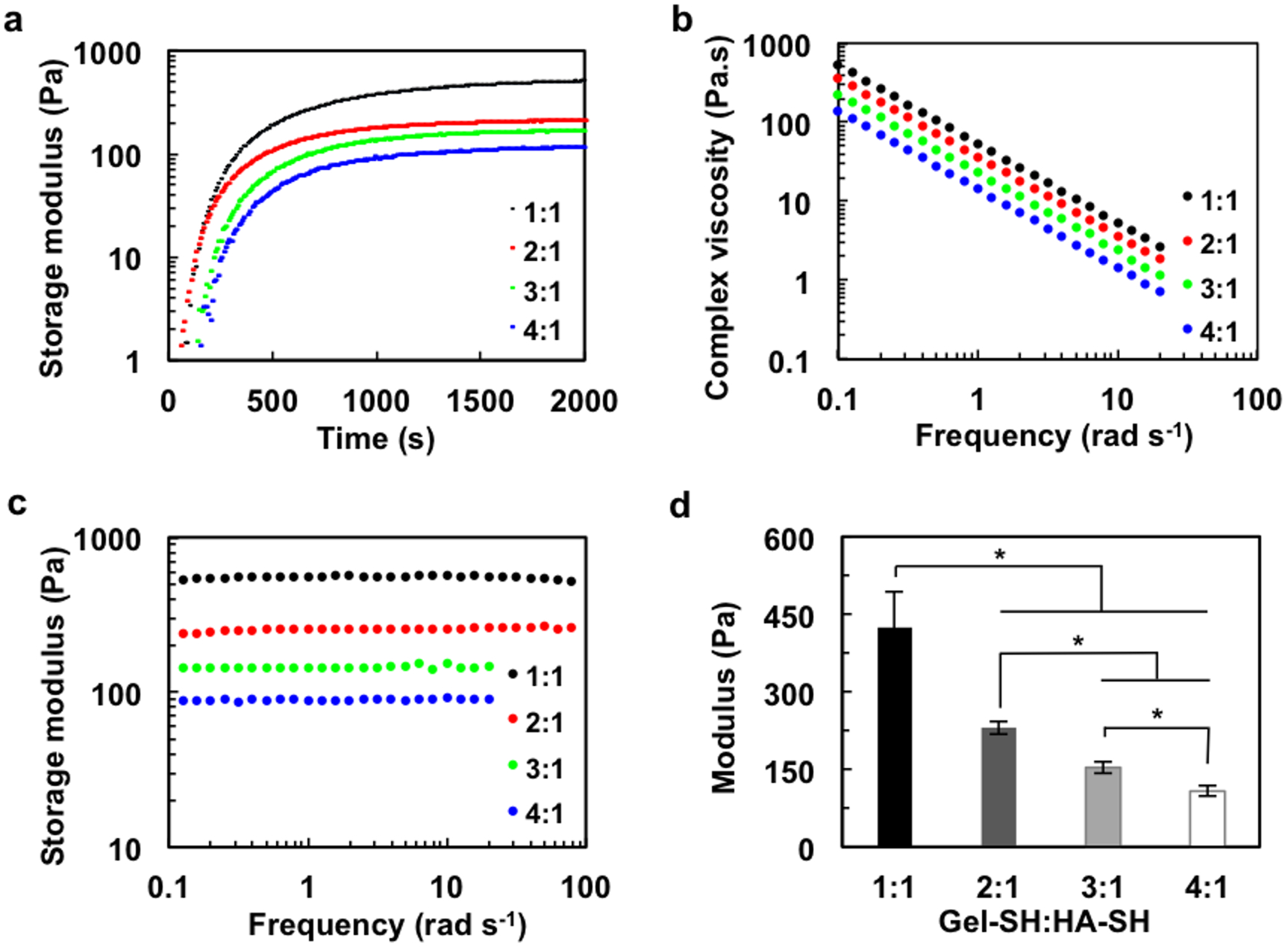
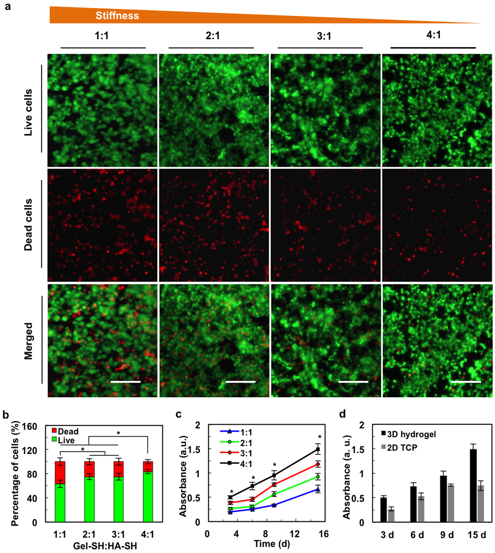
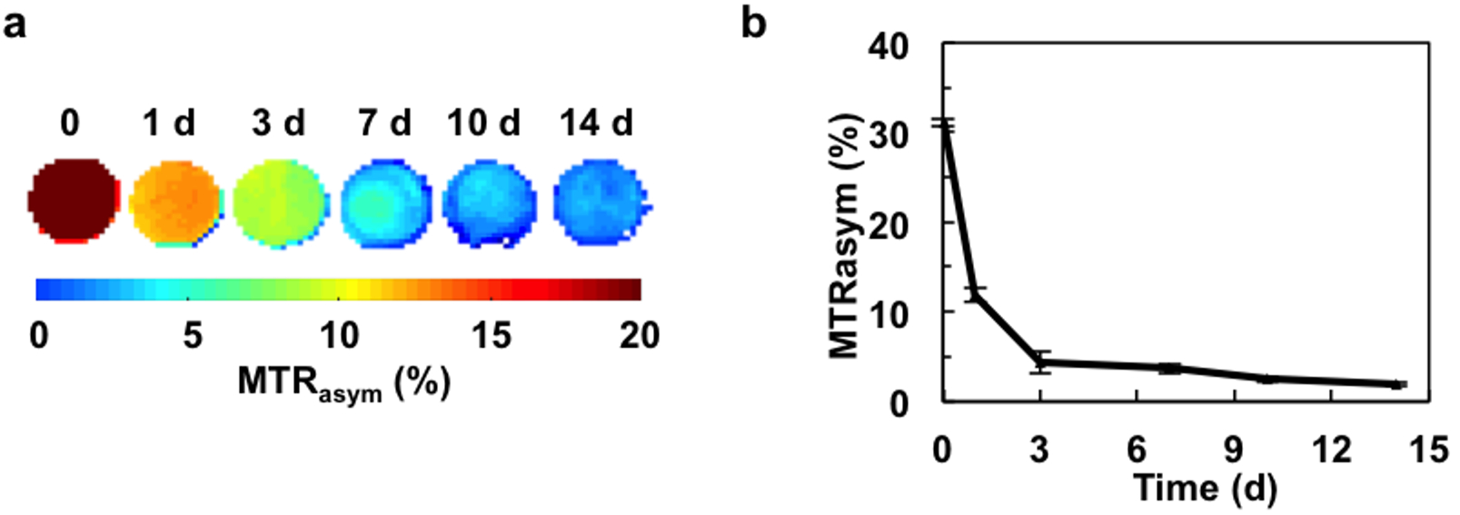
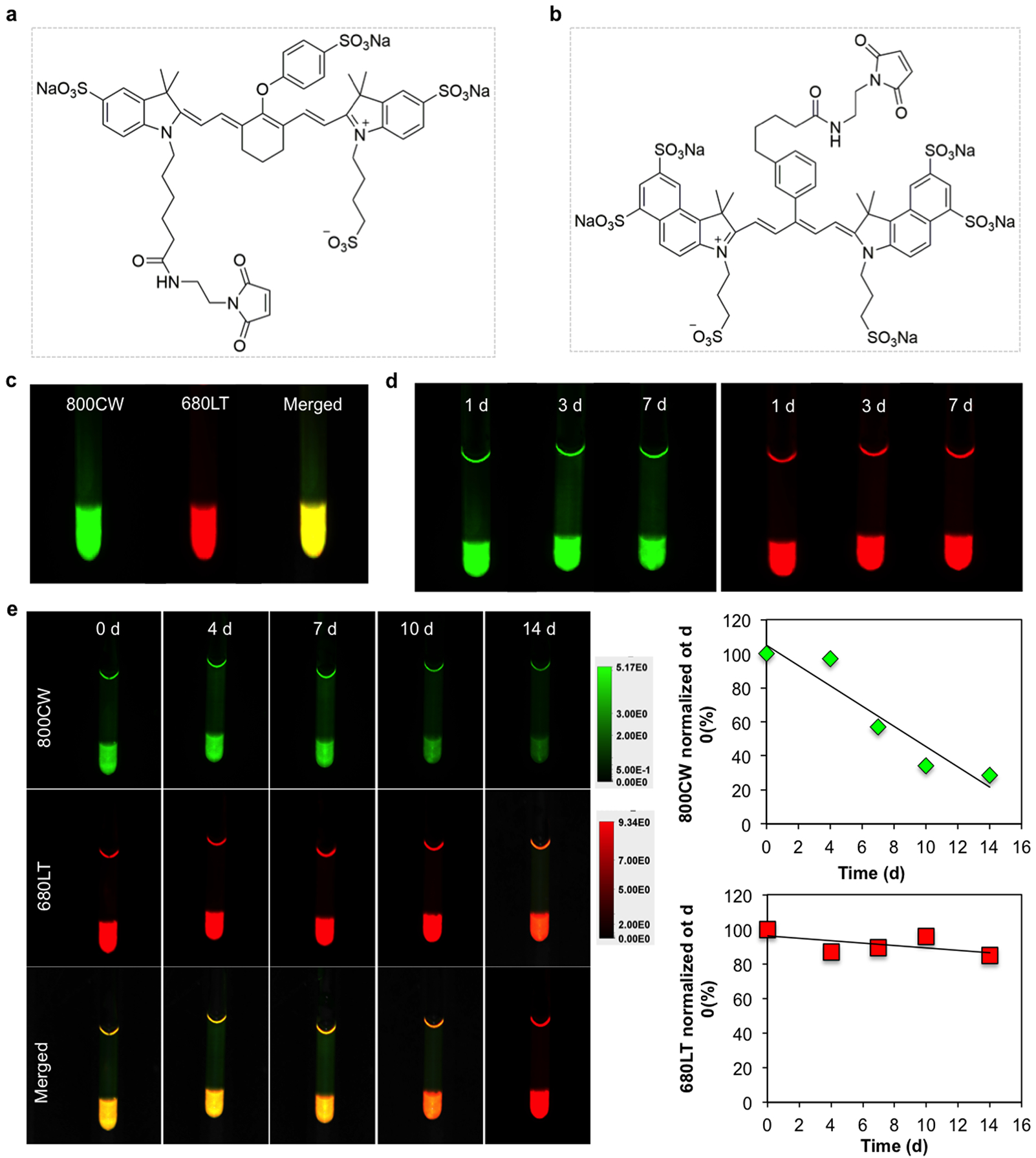
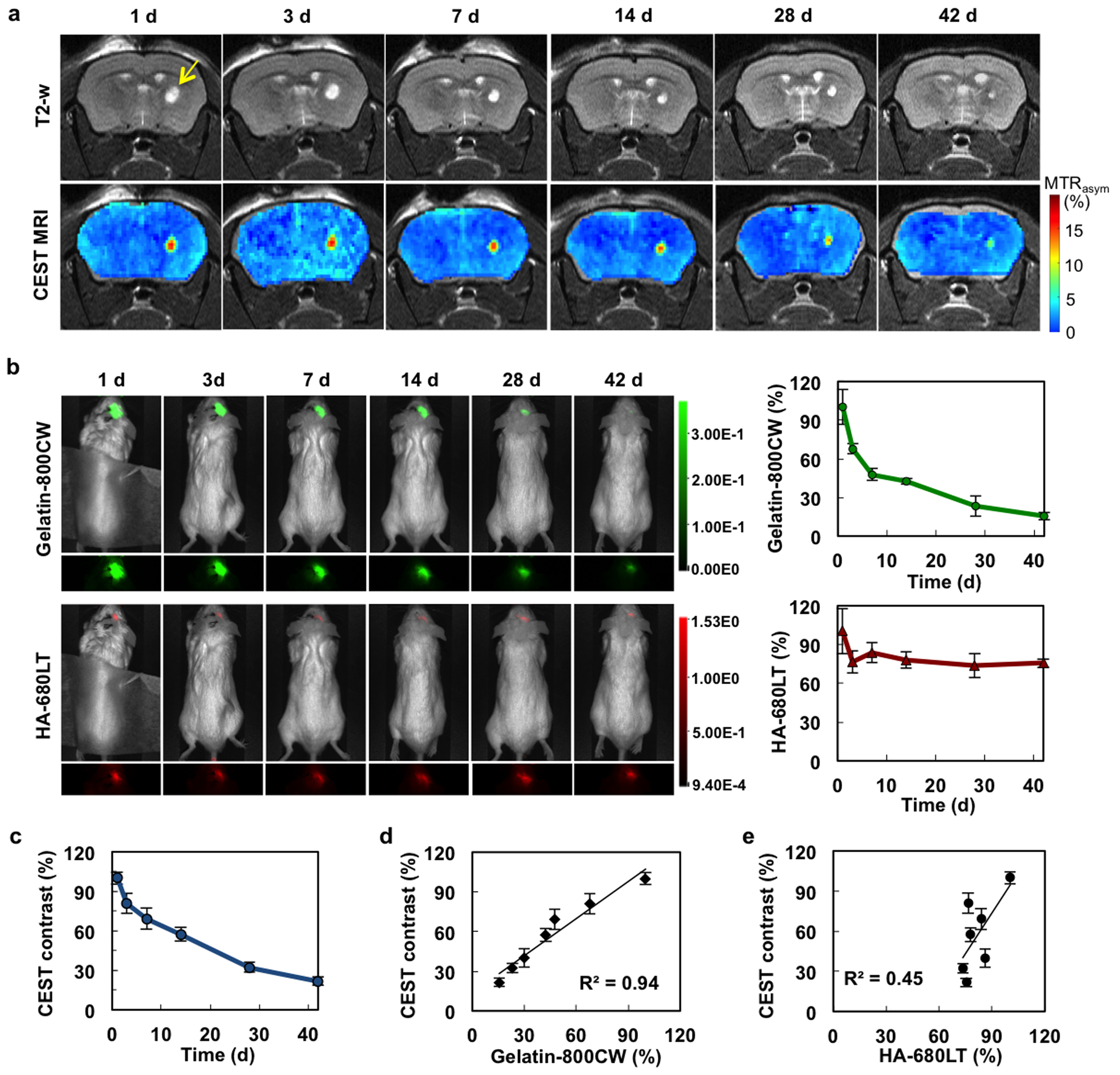
Hydrogel scaffolding of stem cells is a promising strategy to overcome initial cell loss and manipulate cell function post-transplantation. Matrix degradation is a requirement for downstream cell differentiation and functional tissue integration, which determines therapeutic outcome. Therefore, monitoring of hydrogel degradation is essential for scaffolded cell replacement therapies. We show here that chemical exchange saturation transfer magnetic resonance imaging (CEST MRI) can be used as a label-free imaging platform for monitoring the degradation of crosslinked hydrogels containing gelatin (Gel) and hyaluronic acid (HA), of which the stiffness can be fine-tuned by varying the ratio of the Gel:HA. By labeling Gel and HA with two different NIR dyes having distinct emission excitation frequencies, we show here that the HA signal remains stable for 42 days, while the Gel signal gradually decreases to <25% of its initial value at this time point. Both imaging modalities were in excellent agreement for both the time course and relative value of CEST MRI and NIR signals (R=0.94). These findings support the further use of CEST MRI for monitoring biodegradation and optimizing of gelatin-containing hydrogels in a label-free manner.
干细胞水凝胶支架是一种很有前景的策略,可克服移植初期的细胞损失并在移植后调控细胞功能。基质降解是下游细胞分化和功能性组织整合的必要条件,这决定了治疗效果。因此,监测水凝胶降解对于支架细胞替代疗法至关重要。我们在此表明,化学交换饱和转移磁共振成像(CEST MRI)可作为一种无标记成像平台,用于监测含有明胶(Gel)和透明质酸(HA)的交联水凝胶的降解情况,其中水凝胶的硬度可通过改变Gel:HA的比例进行微调。通过用两种具有不同发射激发频率的不同近红外染料标记Gel和HA,我们在此表明,HA信号在42天内保持稳定,而在该时间点Gel信号逐渐降至其初始值的<25%。CEST MRI和近红外信号的时间进程和相对值在两种成像方式中都具有极好的一致性(R = 0.94)。这些发现支持进一步使用CEST MRI以无标记方式监测生物降解并优化含明胶的水凝胶。