,.
.
Invest Ophthalmol Vis Sci. 2020 Mar 9;61(3):35. doi: 10.1167/iovs.61.3.35.
To determine whether parapapillary choroidal microvasculature (PPCMv) density as measured by optical coherence tomography angiography differs between nonarteritic anterior ischemic optic neuropathy (NAION) and primary open angle glaucoma (POAG).
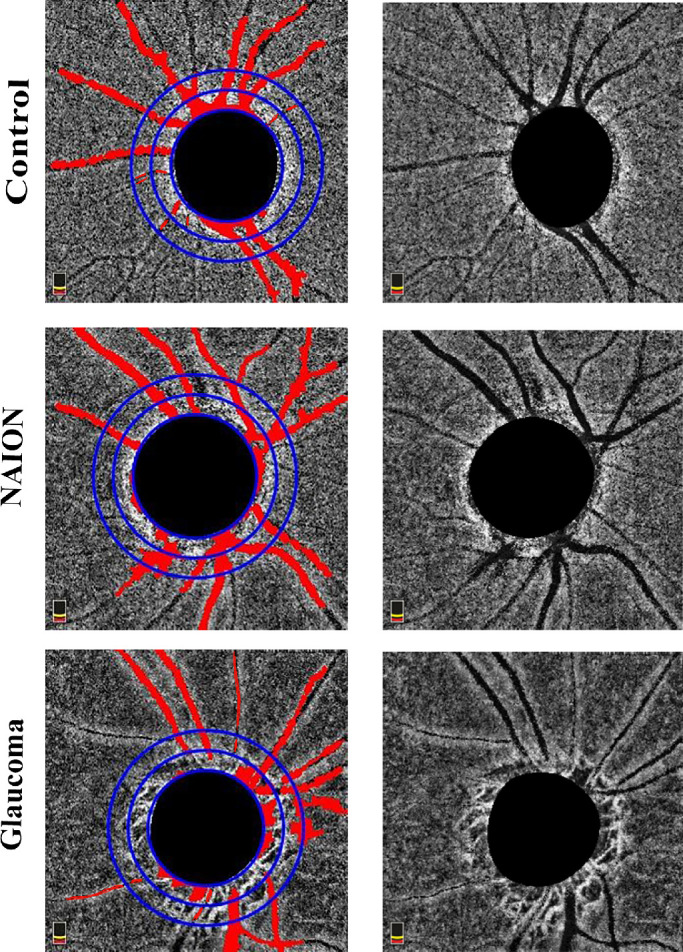
Thirty-seven eyes with chronic NAION, 34 unaffected fellow eyes with NAION, 47 moderate and severe POAG eyes, and 54 healthy control subjects were evaluated. Automated PPCMv density was calculated using custom Matlab software in inner and outer annuli around the optic nerve region in addition to peripapillary superficial retinal vessels.
Linear models showed no difference in peripapillary retinal nerve fiber layer between NAION and POAG eyes. Mean peripapillary superficial small vessels in the NAION and POAG groups were 36.62 ± 7.1% and 39.72 ± 8.18% without a statistically difference between them (P = 0.16). Mean inner and outer annular region PPCMv densities in the NAION group were 26.55 ± 9.2% and 17.81 ± 6.9%, which were not different from unaffected fellow eyes and the control group. However, the POAG group had significantly reduced PPCMv density in both inner and outer annuli with values of 15.84 ± 6.5% and 12.80 ± 5.0%, respectively, compared with normal subjects (both P < 0.001). Inner and outer circle PPCMv densities were also significantly reduced in the POAG group compared with the NAION group.
Reduced PPCMv density in POAG eyes shows that deep optic nerve head ocular blood flow may contribute to axonal damage in patients with glaucoma.
通过光相干断层扫描血管造影术(OCTA)测量视盘旁脉络膜微血管(PPCMv)密度,确定其在非动脉炎性前部缺血性视神经病变(NAION)和原发性开角型青光眼(POAG)之间是否存在差异。
共评估了 37 只患有慢性 NAION 的眼、34 只无 NAION 影响的同眼、47 只中重度 POAG 眼和 54 只健康对照者。使用定制的 Matlab 软件在视神经区域内外环计算自动 PPCMv 密度,此外还计算了视盘周围浅层视网膜血管密度。
线性模型显示,NAION 和 POAG 眼中视盘周围神经纤维层无差异。NAION 和 POAG 组视盘周围浅层小血管平均分别为 36.62 ± 7.1%和 39.72 ± 8.18%,两组间无统计学差异(P = 0.16)。NAION 组内环和外环区域的平均 PPCMv 密度分别为 26.55 ± 9.2%和 17.81 ± 6.9%,与无影响的同眼和对照组无差异。然而,POAG 组内环和外环的 PPCMv 密度均显著降低,分别为 15.84 ± 6.5%和 12.80 ± 5.0%,与正常对照组相比差异均有统计学意义(均 P < 0.001)。与 NAION 组相比,POAG 组内环和外环的 PPCMv 密度也显著降低。
POAG 眼中 PPCMv 密度降低表明,深层视神经头眼血流可能导致青光眼患者轴突损伤。