• 文献检索
  • 文档翻译
  • 深度研究
  • 学术资讯
  • Suppr Zotero 插件Zotero 插件
  • 邀请有礼
  • 套餐&价格
  • 历史记录
应用&插件
Suppr Zotero 插件Zotero 插件浏览器插件Mac 客户端Windows 客户端微信小程序
定价
高级版会员购买积分包购买API积分包
服务
文献检索文档翻译深度研究API 文档MCP 服务
关于我们
关于 Suppr公司介绍联系我们用户协议隐私条款
关注我们

Suppr 超能文献

核心技术专利:CN118964589B侵权必究
粤ICP备2023148730 号-1Suppr @ 2026

文献检索

告别复杂PubMed语法,用中文像聊天一样搜索,搜遍4000万医学文献。AI智能推荐,让科研检索更轻松。

立即免费搜索

文件翻译

保留排版,准确专业,支持PDF/Word/PPT等文件格式,支持 12+语言互译。

免费翻译文档

深度研究

AI帮你快速写综述,25分钟生成高质量综述,智能提取关键信息,辅助科研写作。

立即免费体验

非动脉炎性前部缺血性视神经病变(NAION)患者的光学相干断层扫描血管造影(OCT-A)定量分析与视觉功能相对应。

Quantitative analysis of optical coherence tomographic angiography (OCT-A) in patients with non-arteritic anterior ischemic optic neuropathy (NAION) corresponds to visual function.

机构信息

Department of Ophthalmology, Massachusetts Eye and Ear Infirmary, Harvard Medical School, Boston, MA, United States of America.

Schepens Eye Research Institute, Harvard Medical School, Boston, MA, United States of America.

出版信息

PLoS One. 2018 Jun 28;13(6):e0199793. doi: 10.1371/journal.pone.0199793. eCollection 2018.

DOI:10.1371/journal.pone.0199793
PMID:29953490
原文链接:https://pmc.ncbi.nlm.nih.gov/articles/PMC6023180/
Abstract

PURPOSE

Non-arteritic anterior ischemic optic neuropathy (NAION) is the most common cause of non-glaucomatous optic neuropathy in older adults. Optical coherence tomographic angiography (OCT-A) is an emerging, non-invasive method to study the microvasculature of the posterior pole, including the optic nerve head. The goal of this study was to assess the vascular changes in the optic nerve head and peripapillary area associated with NAION using OCT-A.

DESIGN

Retrospective comparative case series.

METHODS

We performed OCT-A in 25 eyes (7 acute and 18 non-acute) in 19 patients with NAION. Fellow, unaffected eyes were analyzed for comparison. Patent macro- and microvascular densities were quantified in the papillary and peripapillary regions of unaffected, acutely affected, and non-acutely affected eyes and compared across these groups according to laminar segment and capillary sampling region, and with respect to performance on automated visual field testing.

RESULTS

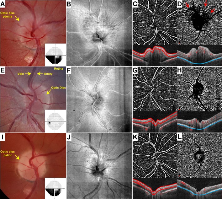
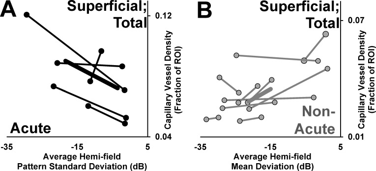
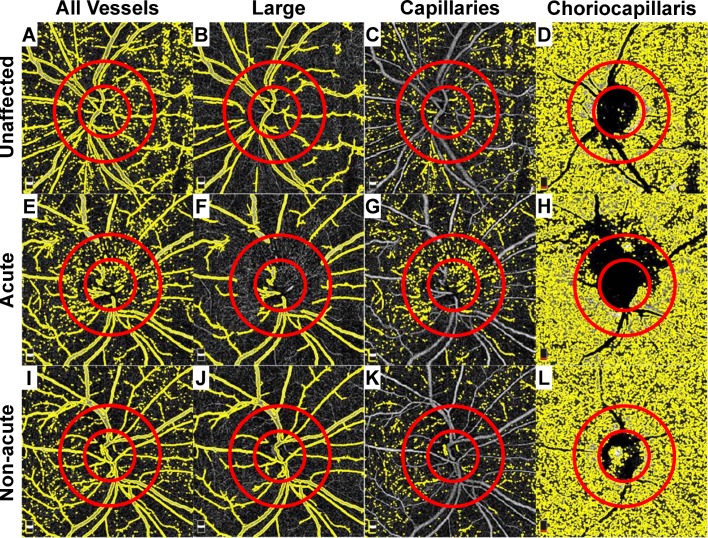
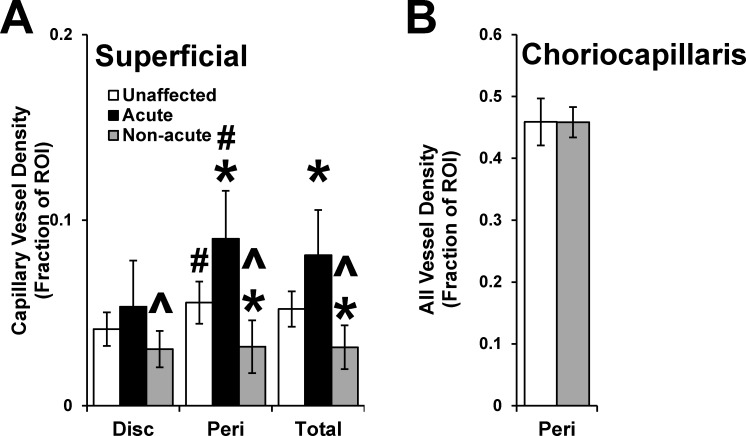
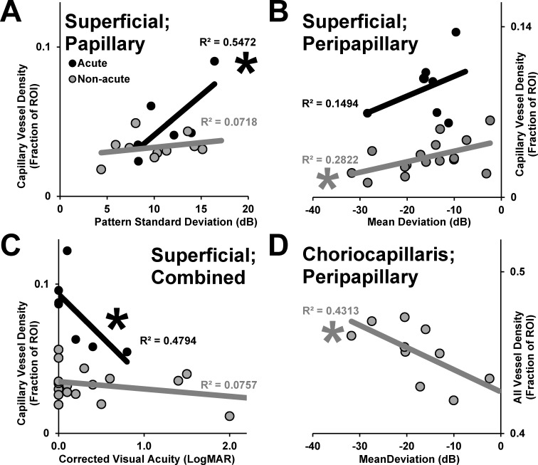
In acutely affected eyes, OCT-A revealed a reduction in the signal from the major retinal vessels and dilation of patent superficial capillaries in the peripapillary area. By contrast, non-acutely affected eyes showed attenuation of patent capillaries. The peripapillary choriocapillaris was obscured by edema in acute cases, but was similar between non-acute and unaffected eyes. The degree of dilation of the superficial microvasculature in the acute phase and attenuation in the non-acute phase each correlated inversely with visual field performance. The region of reduced patent capillary density correlated with the location of visual field defects in 80% of acute cases and 80% of non-acute cases.

CONCLUSIONS

OCT-A reveals a dynamic shift in the superficial capillary network of the optic nerve head with strong functional correlates in both the acute and non-acute phases of NAION. Further study may validate OCT-A as a useful adjunctive diagnostic tool in the evaluation of ischemic optic neuropathy.

摘要

目的

非动脉炎性前部缺血性视神经病变(NAION)是老年人中最常见的非青光眼性视神经病变。光学相干断层血管造影术(OCT-A)是一种新兴的、非侵入性的方法,用于研究后极部的微血管,包括视神经头。本研究的目的是使用 OCT-A 评估与 NAION 相关的视神经头和视盘周围区域的血管变化。

设计

回顾性比较病例系列。

方法

我们对 19 例 NAION 患者的 25 只眼(7 只急性和 18 只非急性)进行了 OCT-A 检查。为了比较,还分析了对侧未受影响的眼睛。在未受影响、急性受影响和非急性受影响的眼中,对乳头和视盘周围区域的显性宏观和微观血管密度进行了量化,并根据层段和毛细血管采样区域对这些组进行了比较,并与自动视野测试的表现进行了比较。

结果

在急性受影响的眼中,OCT-A 显示主要视网膜血管的信号减少,视盘周围区域的显性浅层毛细血管扩张。相比之下,非急性受影响的眼睛显示显性毛细血管衰减。急性病例中,视盘周围脉络膜毛细血管因水肿而模糊,但在非急性和未受影响的眼中相似。在急性期浅层微血管扩张的程度和非急性期的衰减程度均与视野表现呈负相关。显性毛细血管密度降低的区域与 80%的急性病例和 80%的非急性病例的视野缺损部位相关。

结论

OCT-A 显示视神经头浅层毛细血管网络的动态变化,在 NAION 的急性和非急性阶段均与强烈的功能相关性相关。进一步的研究可能会验证 OCT-A 作为评估缺血性视神经病变的有用辅助诊断工具。

https://cdn.ncbi.nlm.nih.gov/pmc/blobs/4efc/6023180/73b890eab6ae/pone.0199793.g005.jpg
https://cdn.ncbi.nlm.nih.gov/pmc/blobs/4efc/6023180/374ad47e8155/pone.0199793.g001.jpg
https://cdn.ncbi.nlm.nih.gov/pmc/blobs/4efc/6023180/46b7e605d516/pone.0199793.g002.jpg
https://cdn.ncbi.nlm.nih.gov/pmc/blobs/4efc/6023180/7faff59f8c4a/pone.0199793.g003.jpg
https://cdn.ncbi.nlm.nih.gov/pmc/blobs/4efc/6023180/097e0ea9aab4/pone.0199793.g004.jpg
https://cdn.ncbi.nlm.nih.gov/pmc/blobs/4efc/6023180/73b890eab6ae/pone.0199793.g005.jpg

相似文献

1
Quantitative analysis of optical coherence tomographic angiography (OCT-A) in patients with non-arteritic anterior ischemic optic neuropathy (NAION) corresponds to visual function.非动脉炎性前部缺血性视神经病变(NAION)患者的光学相干断层扫描血管造影(OCT-A)定量分析与视觉功能相对应。
PLoS One. 2018 Jun 28;13(6):e0199793. doi: 10.1371/journal.pone.0199793. eCollection 2018.
2
Retinal and Choroidal Microvasculature in Nonarteritic Anterior Ischemic Optic Neuropathy: An Optical Coherence Tomography Angiography Study.非动脉炎性前部缺血性视神经病变的视网膜和脉络膜微血管:一项光相干断层扫描血管造影研究。
Invest Ophthalmol Vis Sci. 2018 Feb 1;59(2):870-877. doi: 10.1167/iovs.17-22996.
3
OCT angiography of acute non-arteritic anterior ischemic optic neuropathy.急性非动脉炎性前部缺血性视神经病变的光学相干断层扫描血管造影
J Fr Ophtalmol. 2017 Feb;40(2):102-109. doi: 10.1016/j.jfo.2016.09.020. Epub 2017 Jan 20.
4
Microvascular alterations detected by optical coherence tomography angiography in non-arteritic anterior ischaemic optic neuropathy: a meta-analysis.光学相干断层扫描血管造影术检测非动脉炎性前部缺血性视神经病变中的微血管改变:一项荟萃分析
Acta Ophthalmol. 2022 Mar;100(2):e386-e395. doi: 10.1111/aos.14930. Epub 2021 Jun 21.
5
Microvasculature dropout detected by the optical coherence tomography angiography in nonarteritic anterior ischemic optic neuropathy.光学相干断层扫描血管造影术检测非动脉炎性前部缺血性视神经病变中的微血管缺失
Lasers Surg Med. 2018 Mar;50(3):194-201. doi: 10.1002/lsm.22712. Epub 2017 Oct 7.
6
Optical coherence tomography angiography in acute unilateral nonarteritic anterior ischemic optic neuropathy: A comparison with the fellow eye and with eyes with papilledema.光学相干断层扫描血管造影在急性单侧非动脉炎性前部缺血性视神经病变中的应用:与对侧眼和伴视盘水肿眼的比较。
Indian J Ophthalmol. 2018 Aug;66(8):1144-1148. doi: 10.4103/ijo.IJO_179_18.
7
Optical coherence tomography angiography in acute non-arteritic anterior ischaemic optic neuropathy.光学相干断层扫描血管造影在急性非动脉炎性前部缺血性视神经病变中的应用
Br J Ophthalmol. 2017 Aug;101(8):1045-1051. doi: 10.1136/bjophthalmol-2016-309245. Epub 2017 Jan 5.
8
Optical coherence tomographic angiography identifies peripapillary microvascular dilation and focal non-perfusion in giant cell arteritis.光学相干断层扫描血管造影术可识别巨细胞动脉炎的视盘周围微血管扩张和局灶性无灌注。
Br J Ophthalmol. 2018 Aug;102(8):1141-1146. doi: 10.1136/bjophthalmol-2017-310718. Epub 2017 Nov 9.
9
Comparison of peripapillary retinal nerve fiber layer loss and visual outcome in fellow eyes following sequential bilateral non-arteritic anterior ischemic optic neuropathy.双侧非动脉性前部缺血性视神经病变先后发病后,对侧眼视乳头周围视网膜神经纤维层损失与视觉预后的比较。
Curr Eye Res. 2015 May;40(6):632-7. doi: 10.3109/02713683.2014.952829. Epub 2014 Aug 25.
10
Evaluation of Optic Disc Perfusion with Optical Coherence Tomography Angiography in Acute Non-arteritic Anterior Ischemic Optic Neuropathy.光学相干断层扫描血管造影评估急性非动脉炎性前部缺血性视神经病变的视盘灌注。
Turk J Ophthalmol. 2022 Feb 23;52(1):30-36. doi: 10.4274/tjo.galenos.2021.48465.

引用本文的文献

1
Structural-Functional Correlation in Non-Arteritic Acute Ischemic Optic Neuropathy.非动脉炎性急性缺血性视神经病变中的结构-功能相关性
Eye Brain. 2025 May 3;17:13-25. doi: 10.2147/EB.S512882. eCollection 2025.
2
Impact of Lipid Parameters on Visual Acuity Change in Non-Arteritic Anterior Ischemic Optic Neuropathy.脂质参数对非动脉炎性前部缺血性视神经病变视力变化的影响
Clin Ophthalmol. 2024 Dec 16;18:3791-3800. doi: 10.2147/OPTH.S500368. eCollection 2024.
3
A case-control study of peripapillary microvascular structure by OCT-angiography in non-arteritic ischaemic optic neuropathy at early and resolutive stages.

本文引用的文献

1
Retinal Vessel Density in Optical Coherence Tomography Angiography in Optic Atrophy after Nonarteritic Anterior Ischemic Optic Neuropathy.非动脉性前部缺血性视神经病变后视神经萎缩的光学相干断层扫描血管造影中的视网膜血管密度
J Ophthalmol. 2017;2017:9632647. doi: 10.1155/2017/9632647. Epub 2017 Feb 19.
2
Optical Coherence Tomography Angiography in Nonarteritic Anterior Ischemic Optic Neuropathy.光学相干断层扫描血管造影术在非动脉炎性前部缺血性视神经病变中的应用
J Neuroophthalmol. 2017 Dec;37(4):358-364. doi: 10.1097/WNO.0000000000000493.
3
Optical Coherence Tomography Angiography of the Optic Disc; an Overview.
一项通过光学相干断层扫描血管造影术对非动脉炎性缺血性视神经病变早期和恢复期视乳头周围微血管结构进行的病例对照研究。
Eye (Lond). 2025 Mar;39(4):771-778. doi: 10.1038/s41433-024-03439-w. Epub 2024 Nov 22.
4
Benefit of Optical Coherence Tomography-Angiography in Patients Undergoing Transsphenoidal Pituitary Adenoma Surgery: A Prospective Controlled Study.光学相干断层扫描血管造影术在经蝶窦垂体腺瘤手术患者中的益处:一项前瞻性对照研究。
Diagnostics (Basel). 2024 Aug 12;14(16):1747. doi: 10.3390/diagnostics14161747.
5
Optical coherence tomography angiography findings in patients affected by giant cell arteritis, with and without ocular involvement: a pilot study.巨细胞动脉炎患者伴或不伴眼部受累的光学相干断层扫描血管造影结果:一项初步研究。
Front Med (Lausanne). 2024 Aug 12;11:1408821. doi: 10.3389/fmed.2024.1408821. eCollection 2024.
6
Differential Capillary and Large Vessel Analysis Improves OCTA Classification of Diabetic Retinopathy.差异毛细血管和大血管分析可改善 OCTA 对糖尿病视网膜病变的分类。
Invest Ophthalmol Vis Sci. 2024 Aug 1;65(10):20. doi: 10.1167/iovs.65.10.20.
7
Differential artery-vein analysis improves the OCTA classification of diabetic retinopathy.动静脉差异分析改善了糖尿病视网膜病变的光学相干断层扫描血管造影分类。
Biomed Opt Express. 2024 May 22;15(6):3889-3899. doi: 10.1364/BOE.521657. eCollection 2024 Jun 1.
8
Idiopathic Intracranial Hypertension Papillopathy due to Hormonal Changes during Pregnancy.妊娠期间激素变化所致特发性颅内高压性视乳头病变
Case Rep Ophthalmol Med. 2023 Jul 11;2023:6688445. doi: 10.1155/2023/6688445. eCollection 2023.
9
Vascular Changes in the Macula of Patients after Previous Episodes of Vision Loss Due to Leber Hereditary Optic Neuropathy and Non-Arteritic Ischemic Optic Neuropathy.既往因Leber遗传性视神经病变和非动脉炎性缺血性视神经病变导致视力丧失的患者黄斑区血管变化。
Diagnostics (Basel). 2023 May 12;13(10):1726. doi: 10.3390/diagnostics13101726.
10
An open-source deep learning network AVA-Net for arterial-venous area segmentation in optical coherence tomography angiography.一种用于光学相干断层扫描血管造影中动静脉区域分割的开源深度学习网络AVA-Net。
Commun Med (Lond). 2023 Apr 17;3(1):54. doi: 10.1038/s43856-023-00287-9.
视盘光学相干断层扫描血管造影术概述
J Ophthalmic Vis Res. 2017 Jan-Mar;12(1):98-105. doi: 10.4103/2008-322X.200162.
4
CHOROIDAL THICKNESS IN DIABETIC RETINOPATHY ASSESSED WITH SWEPT-SOURCE OPTICAL COHERENCE TOMOGRAPHY.扫频源光学相干断层扫描评估糖尿病视网膜病变中的脉络膜厚度
Retina. 2018 Jan;38(1):173-182. doi: 10.1097/IAE.0000000000001516.
5
OCT angiography of acute non-arteritic anterior ischemic optic neuropathy.急性非动脉炎性前部缺血性视神经病变的光学相干断层扫描血管造影
J Fr Ophtalmol. 2017 Feb;40(2):102-109. doi: 10.1016/j.jfo.2016.09.020. Epub 2017 Jan 20.
6
Optical coherence tomography angiography in acute non-arteritic anterior ischaemic optic neuropathy.光学相干断层扫描血管造影在急性非动脉炎性前部缺血性视神经病变中的应用
Br J Ophthalmol. 2017 Aug;101(8):1045-1051. doi: 10.1136/bjophthalmol-2016-309245. Epub 2017 Jan 5.
7
SWEPT-SOURCE OPTICAL COHERENCE TOMOGRAPHY ANGIOGRAPHY OF THE OPTIC DISK IN OPTIC NEUROPATHY.视神经病变中视盘的扫频源光学相干断层扫描血管造影
Retina. 2016 Dec;36 Suppl 1:S168-S177. doi: 10.1097/IAE.0000000000001259.
8
Structural and Functional Analyses in Nonarteritic Anterior Ischemic Optic Neuropathy: Optical Coherence Tomography Angiography Study.非动脉炎性前部缺血性视神经病变的结构与功能分析:光学相干断层扫描血管造影研究
J Neuroophthalmol. 2017 Jun;37(2):140-148. doi: 10.1097/WNO.0000000000000470.
9
Optical Coherence Tomography Angiography in a Patient with Optic Atrophy After Non-arteritic Anterior Ischaemic Optic Neuropathy.非动脉炎性前部缺血性视神经病变后视神经萎缩患者的光学相干断层扫描血管造影
Neuroophthalmology. 2016 Apr 4;40(3):146-149. doi: 10.3109/01658107.2016.1162174. eCollection 2016 Jun.
10
Clinical Usefulness of Spectral-Domain Optical Coherence Tomography in Glaucoma and NAION.频域光学相干断层扫描在青光眼和非动脉炎性前部缺血性视神经病变中的临床应用价值
Chonnam Med J. 2016 Sep;52(3):194-200. doi: 10.4068/cmj.2016.52.3.194. Epub 2016 Sep 23.