• 文献检索
  • 文档翻译
  • 深度研究
  • 学术资讯
  • Suppr Zotero 插件Zotero 插件
  • 邀请有礼
  • 套餐&价格
  • 历史记录
应用&插件
Suppr Zotero 插件Zotero 插件浏览器插件Mac 客户端Windows 客户端微信小程序
定价
高级版会员购买积分包购买API积分包
服务
文献检索文档翻译深度研究API 文档MCP 服务
关于我们
关于 Suppr公司介绍联系我们用户协议隐私条款
关注我们

Suppr 超能文献

核心技术专利:CN118964589B侵权必究
粤ICP备2023148730 号-1Suppr @ 2026

文献检索

告别复杂PubMed语法,用中文像聊天一样搜索,搜遍4000万医学文献。AI智能推荐,让科研检索更轻松。

立即免费搜索

文件翻译

保留排版,准确专业,支持PDF/Word/PPT等文件格式,支持 12+语言互译。

免费翻译文档

深度研究

AI帮你快速写综述,25分钟生成高质量综述,智能提取关键信息,辅助科研写作。

立即免费体验

与原发性开角型青光眼相比,缺血性视神经病变中视乳头周围毛细血管密度丧失的模式。

Pattern of peripapillary capillary density loss in ischemic optic neuropathy compared to that in primary open-angle glaucoma.

作者信息

Fard Masoud Aghsaei, Suwan Yanin, Moghimi Sasan, Geyman Lawrence S, Chui Toco Y, Rosen Richard B, Ritch Robert

机构信息

Farabi Eye Hospital, Tehran University of Medical Science, Tehran, Iran.

Department of Ophthalmology, Ramathibodi Hospital, Mahidol University, Bangkok, Thailand.

出版信息

PLoS One. 2018 Jan 10;13(1):e0189237. doi: 10.1371/journal.pone.0189237. eCollection 2018.

DOI:10.1371/journal.pone.0189237
PMID:29320503
原文链接:https://pmc.ncbi.nlm.nih.gov/articles/PMC5761857/
Abstract

PURPOSE

Both non-arteritic anterior ischemic optic neuropathy (NAION) and primary open-angle glaucoma (POAG) damage retinal ganglion cell axons, which are perfused by the radial peripapillary capillaries. To evaluate the pattern of ischemia, we compared peripapillary capillary density (PCD) in NAION eyes to POAG eyes matched for visual field mean deviation and retinal nerve fiber layer thickness.

METHODS

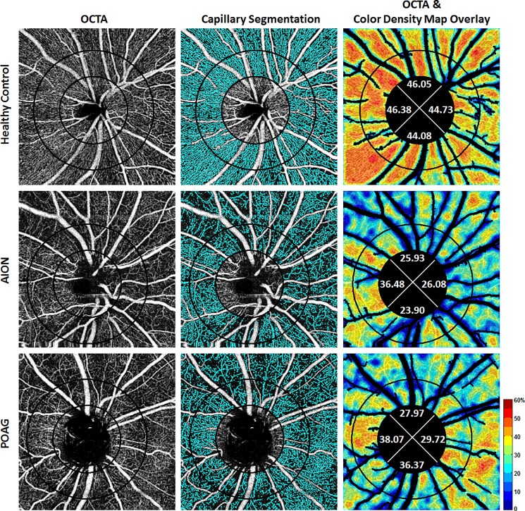
31 chronic NAION (>6 months after the acute event) and unaffected fellow eyes (31 subjects), 42 moderate and severe POAG eyes (27 subjects), and 77 control eyes (46 healthy subjects) were imaged with a commercial optical coherence tomography angiography system (AngioVue, Avanti RTVue-XR, Optovue, CA) at two academic institutions. Two concentric circles of diameters 1.95mm (inner) and 3.45mm (outer) were manually placed on images centered on the optic nerve head, producing an annular region-of-interest. Image analysis with major vessel removal was performed using a custom program. Whole-image, whole-annulus, and sectoral PCDs were measured.

RESULTS

Whole-image and whole-annulus PCDs in NAION and moderate and severe POAG eyes were significantly decreased compared to unaffected fellow eyes and control eyes (all P<0.001). Superior and temporal PCD values were affected more than other sectors in both NAION and POAG groups compared to control group. Whole-image and whole-annulus PCDs were not statistically different between NAION and POAG eyes (both P = 0.99). However, of all peripapillary sectors, the inferior sector PCD value was less affected in POAG eyes compared to NAION eyes (P = 0.001). Univariate analysis results also revealed a significant positive correlation between superior and inferior PCDs and corresponding RNFL thicknesses. The inferior sector correlation was greater in POAG than NAION eyes.

CONCLUSION

While the whole PCD values were not different in chronic NAION and POAG, the greater correlation of inferior PCD with corresponding RNFL sectors in POAG compared to NAION suggests greater susceptibility of the inferior radial peripapillary capillary in the pathogenesis of POAG.

摘要

目的

非动脉炎性前部缺血性视神经病变(NAION)和原发性开角型青光眼(POAG)都会损害由视乳头周围放射状毛细血管灌注的视网膜神经节细胞轴突。为了评估缺血模式,我们比较了NAION患眼与视野平均偏差和视网膜神经纤维层厚度相匹配的POAG患眼的视乳头周围毛细血管密度(PCD)。

方法

在两家学术机构,使用商用光学相干断层扫描血管造影系统(AngioVue,Avanti RTVue-XR,Optovue,加利福尼亚州)对31例慢性NAION患者(急性事件发生后>6个月)及其未受影响的对侧眼(31名受试者)、42例中重度POAG患眼(27名受试者)和77只对照眼(46名健康受试者)进行成像。在以视神经乳头为中心的图像上手动放置两个直径分别为1.95mm(内)和3.45mm(外)的同心圆,形成一个环形感兴趣区域。使用定制程序进行去除主要血管后的图像分析。测量全图像、全环和扇形PCD。

结果

与未受影响的对侧眼和对照眼相比,NAION患眼以及中重度POAG患眼的全图像和全环PCD均显著降低(所有P<0.001)。与对照组相比,NAION组和POAG组的上方和颞侧PCD值受影响程度大于其他扇形区域。NAION患眼和POAG患眼的全图像和全环PCD在统计学上无差异(两者P = 0.99)。然而,在所有视乳头周围扇形区域中,与NAION患眼相比,POAG患眼的下方扇形区域PCD值受影响较小(P = 0.001)。单因素分析结果还显示上方和下方PCD与相应的视网膜神经纤维层厚度之间存在显著正相关。POAG患眼下方扇形区域的相关性大于NAION患眼。

结论

虽然慢性NAION和POAG的整体PCD值没有差异,但与NAION相比,POAG中下方PCD与相应视网膜神经纤维层扇形区域的相关性更高,这表明在POAG发病机制中,下方视乳头周围放射状毛细血管的易损性更大。

https://cdn.ncbi.nlm.nih.gov/pmc/blobs/2662/5761857/d1a6a93f5125/pone.0189237.g001.jpg

相似文献

1
Pattern of peripapillary capillary density loss in ischemic optic neuropathy compared to that in primary open-angle glaucoma.与原发性开角型青光眼相比,缺血性视神经病变中视乳头周围毛细血管密度丧失的模式。
PLoS One. 2018 Jan 10;13(1):e0189237. doi: 10.1371/journal.pone.0189237. eCollection 2018.
2
Comparison of the Pattern of Macular Ganglion Cell-Inner Plexiform Layer Defect Between Ischemic Optic Neuropathy and Open-Angle Glaucoma.缺血性视神经病变与开角型青光眼黄斑神经节细胞-内层神经纤维层缺损模式的比较
Invest Ophthalmol Vis Sci. 2016 Mar;57(3):1011-6. doi: 10.1167/iovs.15-18618.
3
Microvascular and structural alterations in the optic nerve head of advanced primary open-angle glaucoma compared with atrophic non-arteritic anterior ischemic optic neuropathy.与非动脉炎性前部缺血性视神经病变相比,晚期原发性开角型青光眼的视神经头微血管和结构改变。
Graefes Arch Clin Exp Ophthalmol. 2021 Jul;259(7):1945-1953. doi: 10.1007/s00417-021-05122-2. Epub 2021 Mar 4.
4
Peripapillary Perfused Capillary Density in Exfoliation Syndrome and Exfoliation Glaucoma versus POAG and Healthy Controls: An OCTA Study.剥脱综合征和剥脱性青光眼与原发性开角型青光眼及健康对照者的视乳头周围灌注毛细血管密度:一项光学相干断层扫描血管造影研究
Asia Pac J Ophthalmol (Phila). 2018 Mar-Apr;7(2):84-89. doi: 10.22608/APO.2017318. Epub 2017 Nov 22.
5
Difference in patterns of retinal ganglion cell damage between primary open-angle glaucoma and non-arteritic anterior ischaemic optic neuropathy.原发性开角型青光眼与非动脉性前部缺血性视神经病变视网膜神经节细胞损伤模式的差异
PLoS One. 2017 Oct 26;12(10):e0187093. doi: 10.1371/journal.pone.0187093. eCollection 2017.
6
Automated Evaluation of Parapapillary Choroidal Microvasculature in Ischemic Optic Neuropathy and Open Angle Glaucoma.缺血性视神经病变和开角型青光眼的视盘旁脉络膜微循环的自动评估
Invest Ophthalmol Vis Sci. 2020 Mar 9;61(3):35. doi: 10.1167/iovs.61.3.35.
7
Macular Vascularity in Ischemic Optic Neuropathy Compared to Glaucoma by Projection-Resolved Optical Coherence Tomography Angiography.基于层析分辨光学相干断层血管造影术的缺血性视神经病变与青光眼的黄斑血管密度比较。
Am J Ophthalmol. 2020 Jan;209:27-34. doi: 10.1016/j.ajo.2019.09.015. Epub 2019 Sep 25.
8
Optic Nerve Head Morphology in Nonarteritic Anterior Ischemic Optic Neuropathy Compared to Open-Angle Glaucoma.非动脉炎性前部缺血性视神经病变与开角型青光眼的视神经乳头形态学比较
Invest Ophthalmol Vis Sci. 2016 Sep 1;57(11):4632-40. doi: 10.1167/iovs.16-19442.
9
Optical Coherence Tomography Angiography Analysis of Perfused Peripapillary Capillaries in Primary Open-Angle Glaucoma and Normal-Tension Glaucoma.原发性开角型青光眼和正常眼压性青光眼视乳头周围灌注毛细血管的光学相干断层扫描血管造影分析
Invest Ophthalmol Vis Sci. 2016 Jul 1;57(9):OCT611-OCT620. doi: 10.1167/iovs.15-18945.
10
Comparison of peripapillary retinal nerve fiber layer loss and visual outcome in fellow eyes following sequential bilateral non-arteritic anterior ischemic optic neuropathy.双侧非动脉性前部缺血性视神经病变先后发病后,对侧眼视乳头周围视网膜神经纤维层损失与视觉预后的比较。
Curr Eye Res. 2015 May;40(6):632-7. doi: 10.3109/02713683.2014.952829. Epub 2014 Aug 25.

引用本文的文献

1
Quantitative assessment of retinal microvasculature using optical coherence tomography angiography and correlation with visual acuity in leber's hereditary optic neuropathy.使用光学相干断层扫描血管造影术对Leber遗传性视神经病变患者视网膜微血管进行定量评估及其与视力的相关性研究
Int Ophthalmol. 2025 Sep 6;45(1):376. doi: 10.1007/s10792-025-03619-x.
2
Optical coherence tomography angiography findings in patients affected by giant cell arteritis, with and without ocular involvement: a pilot study.巨细胞动脉炎患者伴或不伴眼部受累的光学相干断层扫描血管造影结果:一项初步研究。
Front Med (Lausanne). 2024 Aug 12;11:1408821. doi: 10.3389/fmed.2024.1408821. eCollection 2024.
3

本文引用的文献

1
Comparison of the Retinal Microvascular Density Between Open Angle Glaucoma and Nonarteritic Anterior Ischemic Optic Neuropathy.开角型青光眼与非动脉性前部缺血性视神经病变视网膜微血管密度的比较。
Invest Ophthalmol Vis Sci. 2017 Jul 1;58(9):3350-3356. doi: 10.1167/iovs.17-22021.
2
Peripapillary Retinal Nerve Fiber Layer Vascular Microcirculation in Eyes With Glaucoma and Single-Hemifield Visual Field Loss.青光眼伴单眼半视野缺损患者的视乳头周围视网膜神经纤维层血管微循环
JAMA Ophthalmol. 2017 May 1;135(5):461-468. doi: 10.1001/jamaophthalmol.2017.0261.
3
Optical Coherence Tomography Angiography in Nonarteritic Anterior Ischemic Optic Neuropathy.
Peripapillary Vascular Density in Compressive Optic Neuropathy and Normal-Tension Glaucoma: A Severity-Controlled Comparison.
压迫性视神经病变和正常眼压性青光眼的视盘旁血管密度:严重程度对照比较。
Invest Ophthalmol Vis Sci. 2023 Sep 1;64(12):10. doi: 10.1167/iovs.64.12.10.
4
Disagreement of Radial Peripapillary Capillary Density Among Four Optical Coherence Tomography Angiography Devices.四种光学相干断层扫描血管造影设备的视盘周围毛细血管密度存在差异。
Transl Vis Sci Technol. 2023 Aug 1;12(8):7. doi: 10.1167/tvst.12.8.7.
5
Peripapillary and macular microvasculature features of non-arteritic anterior ischemic optic neuropathy.非动脉炎性前部缺血性视神经病变的视乳头周围和黄斑部微血管特征
Front Med (Lausanne). 2023 Jan 12;9:1033838. doi: 10.3389/fmed.2022.1033838. eCollection 2022.
6
A Deep Learning System Using Optical Coherence Tomography Angiography to Detect Glaucoma and Anterior Ischemic Optic Neuropathy.一种使用光学相干断层扫描血管造影术检测青光眼和前部缺血性视神经病变的深度学习系统。
J Clin Med. 2023 Jan 7;12(2):507. doi: 10.3390/jcm12020507.
7
The Role of Optical Coherence Tomography Angiography in Optic Nerve Head Edema: A Narrative Review.光学相干断层扫描血管造影在视神经乳头水肿中的作用:一项叙述性综述
J Ophthalmol. 2022 Nov 30;2022:5823345. doi: 10.1155/2022/5823345. eCollection 2022.
8
Optical coherence tomography evaluation of retinal nerve fiber layer thickness in non-arteritic anterior ischemic optic neuropathy and primary open angle glaucoma: a systematic review and Meta-analysis.光学相干断层扫描评估非动脉炎性前部缺血性视神经病变和原发性开角型青光眼中视网膜神经纤维层厚度:一项系统评价和Meta分析。
Int J Ophthalmol. 2022 Aug 18;15(8):1370-1380. doi: 10.18240/ijo.2022.08.22. eCollection 2022.
9
Reduced choroidal peripapillary capillaries in thyroid-associated ophthalmopathy with early stage of dysthyroid optic neuropathy.甲状腺相关性眼病伴甲状腺功能异常性视神经病变早期脉络膜视乳头周围毛细血管减少
Int J Ophthalmol. 2022 Jul 18;15(7):1135-1141. doi: 10.18240/ijo.2022.07.14. eCollection 2022.
10
A Pilot Study of Subclinical Non-Capillary Peripapillary Perfusion Changes in Thyroid-Related Orbitopathy Detected Using Optical Coherence Tomography Angiography.一项利用光学相干断层扫描血管造影术检测甲状腺相关性眼病亚临床非毛细血管视乳头周围灌注变化的初步研究。
Clin Ophthalmol. 2022 Mar 20;16:867-875. doi: 10.2147/OPTH.S356631. eCollection 2022.
光学相干断层扫描血管造影术在非动脉炎性前部缺血性视神经病变中的应用
J Neuroophthalmol. 2017 Dec;37(4):358-364. doi: 10.1097/WNO.0000000000000493.
4
Peripapillary perfused capillary density in primary openangle glaucoma across disease stage an optical coherence tomography angiography study.原发性开角型青光眼不同疾病阶段的视乳头周围灌注毛细血管密度:一项光学相干断层扫描血管造影研究
Br J Ophthalmol. 2017 Sep;101(9):1261-1268. doi: 10.1136/bjophthalmol-2016-309642. Epub 2017 Feb 1.
5
Structural and Functional Analyses in Nonarteritic Anterior Ischemic Optic Neuropathy: Optical Coherence Tomography Angiography Study.非动脉炎性前部缺血性视神经病变的结构与功能分析:光学相干断层扫描血管造影研究
J Neuroophthalmol. 2017 Jun;37(2):140-148. doi: 10.1097/WNO.0000000000000470.
6
OCT Angiography of the Peripapillary Retina in Primary Open-Angle Glaucoma.原发性开角型青光眼视乳头周围视网膜的光学相干断层扫描血管造影
Invest Ophthalmol Vis Sci. 2016 Nov 1;57(14):6265-6270. doi: 10.1167/iovs.16-20287.
7
Optical Coherence Tomography Angiography Analysis of Perfused Peripapillary Capillaries in Primary Open-Angle Glaucoma and Normal-Tension Glaucoma.原发性开角型青光眼和正常眼压性青光眼视乳头周围灌注毛细血管的光学相干断层扫描血管造影分析
Invest Ophthalmol Vis Sci. 2016 Jul 1;57(9):OCT611-OCT620. doi: 10.1167/iovs.15-18945.
8
Relationship between Optical Coherence Tomography Angiography Vessel Density and Severity of Visual Field Loss in Glaucoma.光学相干断层扫描血管造影血管密度与青光眼视野缺损严重程度之间的关系
Ophthalmology. 2016 Dec;123(12):2498-2508. doi: 10.1016/j.ophtha.2016.08.041. Epub 2016 Oct 7.
9
Quantitative Optical Coherence Tomography Angiography of Radial Peripapillary Capillaries in Glaucoma, Glaucoma Suspect, and Normal Eyes.青光眼、疑似青光眼和正常眼的视盘周围放射状毛细血管的定量光学相干断层扫描血管造影
Am J Ophthalmol. 2016 Oct;170:41-49. doi: 10.1016/j.ajo.2016.07.015. Epub 2016 Jul 25.
10
Optical Coherence Tomography Angiography Vessel Density in Healthy, Glaucoma Suspect, and Glaucoma Eyes.健康眼、青光眼可疑眼和青光眼眼中光学相干断层扫描血管造影的血管密度
Invest Ophthalmol Vis Sci. 2016 Jul 1;57(9):OCT451-9. doi: 10.1167/iovs.15-18944.