• 文献检索
  • 文档翻译
  • 深度研究
  • 学术资讯
  • Suppr Zotero 插件Zotero 插件
  • 邀请有礼
  • 套餐&价格
  • 历史记录
应用&插件
Suppr Zotero 插件Zotero 插件浏览器插件Mac 客户端Windows 客户端微信小程序
定价
高级版会员购买积分包购买API积分包
服务
文献检索文档翻译深度研究API 文档MCP 服务
关于我们
关于 Suppr公司介绍联系我们用户协议隐私条款
关注我们

Suppr 超能文献

核心技术专利:CN118964589B侵权必究
粤ICP备2023148730 号-1Suppr @ 2026

文献检索

告别复杂PubMed语法,用中文像聊天一样搜索,搜遍4000万医学文献。AI智能推荐,让科研检索更轻松。

立即免费搜索

文件翻译

保留排版,准确专业,支持PDF/Word/PPT等文件格式,支持 12+语言互译。

免费翻译文档

深度研究

AI帮你快速写综述,25分钟生成高质量综述,智能提取关键信息,辅助科研写作。

立即免费体验

术中 CT 和锥形束 CT 成像在自发性脑出血微创清除术中的应用。

Intraoperative CT and cone-beam CT imaging for minimally invasive evacuation of spontaneous intracerebral hemorrhage.

机构信息

Department of Neurosurgery and Center for Stroke Research Berlin (CSB), Charité - Universitätsmedizin Berlin, Charitéplatz 1, 10117, Berlin, Germany.

Department of Neuroradiology, Charité - Universitätsmedizin Berlin, Berlin, Germany.

出版信息

Acta Neurochir (Wien). 2020 Dec;162(12):3167-3177. doi: 10.1007/s00701-020-04284-y. Epub 2020 Mar 19.

DOI:10.1007/s00701-020-04284-y
PMID:32193726
原文链接:https://pmc.ncbi.nlm.nih.gov/articles/PMC7593312/
Abstract

BACKGROUND

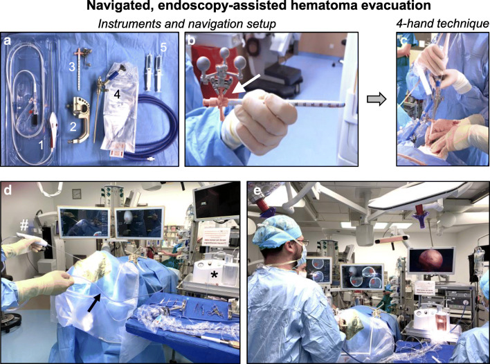
Minimally invasive surgery (MIS) for evacuation of spontaneous intracerebral hemorrhage (ICH) has shown promise but there remains a need for intraoperative performance assessment considering the wide range of evacuation effectiveness. In this feasibility study, we analyzed the benefit of intraoperative 3-dimensional imaging during navigated endoscopy-assisted ICH evacuation by mechanical clot fragmentation and aspiration.

METHODS

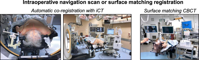
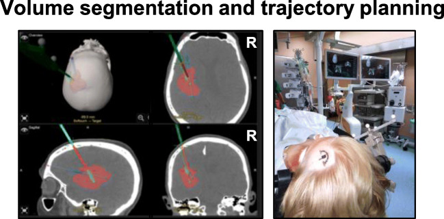
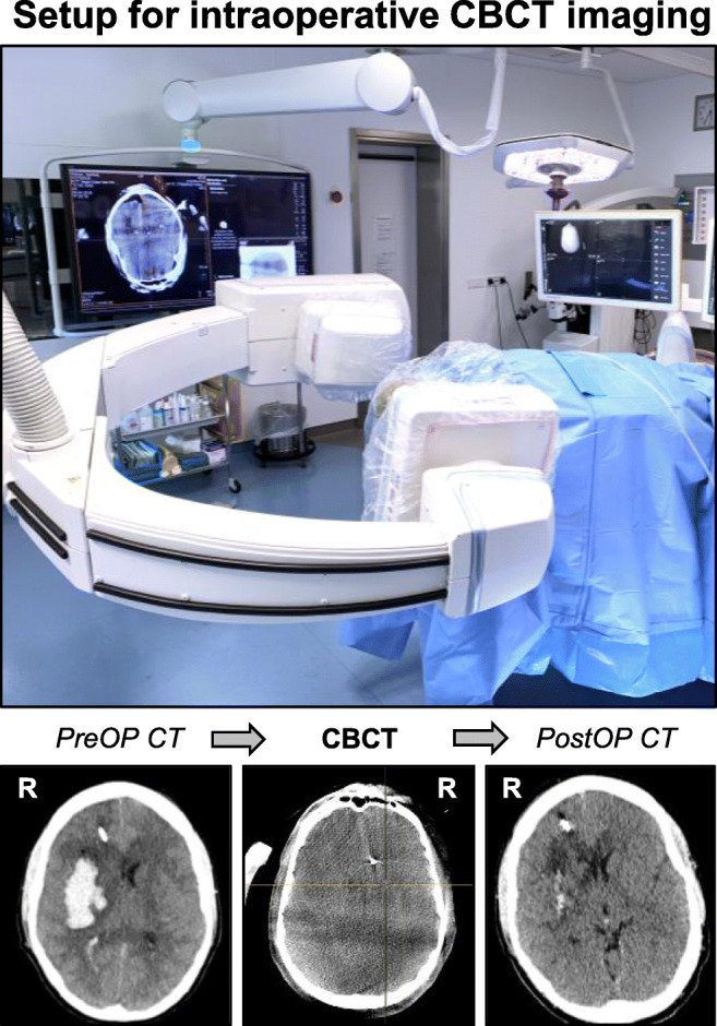
18 patients with superficial or deep supratentorial ICH underwent MIS for clot evacuation followed by intraoperative computerized tomography (iCT) or cone-beam CT (CBCT) imaging. Eligibility for MIS required (a) availability of intraoperative iCT or CBCT, (b) spontaneous lobar or deep ICH without vascular pathology, (c) a stable ICH volume (20-90 ml), (d) a reduced level of consciousness (GCS 5-14), and (e) a premorbid mRS ≤ 1. Demographic, clinical, and radiographic patient data were analyzed by two independent observers.

RESULTS

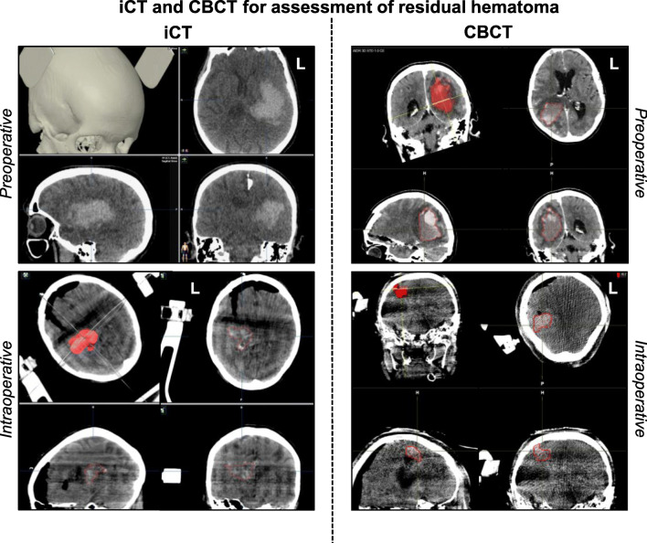
Nine female and 9 male patients with a median age of 76 years (42-85) presented with an ICH score of 3 (1-4), GCS of 10 (5-14) and ICH volume of 54 ± 26 ml. Clot fragmentation and aspiration was feasible in all cases and intraoperative imaging determined an overall evacuation rate of 80 ± 19% (residual hematoma volume: 13 ± 17 ml; p < 0.0001 vs. Pre-OP). Based on the intraoperative imaging results, 1/3rd of all patients underwent an immediate re-aspiration attempt. No patient experienced hemorrhagic complications or required conversion to open craniotomy. However, routine postoperative CT imaging revealed early hematoma re-expansion with an adjusted evacuation rate of 59 ± 30% (residual hematoma volume: 26 ± 37 ml; p < 0.001 vs. Pre-OP).

CONCLUSIONS

Routine utilization of iCT or CBCT imaging in MIS for ICH permits direct surgical performance assessment and the chance for immediate re-aspiration, which may optimize targeting of an ideal residual hematoma volume and reduce secondary revision rates.

摘要

背景

微创血肿清除术(MIS)治疗自发性脑出血(ICH)显示出了良好的效果,但考虑到清除效果的广泛差异,仍需要对术中操作进行评估。在这项可行性研究中,我们分析了导航内镜辅助机械性血栓碎裂抽吸清除ICH 过程中术中三维成像的作用。

方法

18 例幕上浅部或深部 ICH 患者接受了 MIS 血肿清除术,之后进行术中计算机断层扫描(iCT)或锥形束 CT(CBCT)成像。MIS 的入选标准为:(a)术中可行 iCT 或 CBCT,(b)无血管病变的自发性脑叶或深部 ICH,(c)血肿体积稳定(20-90ml),(d)意识水平下降(GCS 5-14),(e)发病前 mRS 评分≤1。两名独立观察者对患者的人口统计学、临床和影像学数据进行了分析。

结果

9 例女性和 9 例男性患者,年龄中位数为 76 岁(42-85 岁),ICH 评分 3 分(1-4 分),GCS 评分 10 分(5-14 分),ICH 体积 54±26ml。所有患者均可行血栓碎裂和抽吸术,术中成像确定总体清除率为 80±19%(残余血肿体积:13±17ml;p<0.0001 与术前相比)。根据术中成像结果,1/3 的患者立即进行了再次抽吸尝试。无患者发生出血性并发症或需要转为开颅手术。然而,常规术后 CT 成像显示早期血肿再扩张,调整后的清除率为 59±30%(残余血肿体积:26±37ml;p<0.001 与术前相比)。

结论

在 MIS 治疗 ICH 中常规使用 iCT 或 CBCT 成像可直接进行手术操作评估,并可立即进行再次抽吸,这可能有助于确定理想的残余血肿体积,并降低二次修正率。

https://cdn.ncbi.nlm.nih.gov/pmc/blobs/362a/7593312/948936a9a2ae/701_2020_4284_Fig6_HTML.jpg
https://cdn.ncbi.nlm.nih.gov/pmc/blobs/362a/7593312/c7ec99955f6e/701_2020_4284_Fig1_HTML.jpg
https://cdn.ncbi.nlm.nih.gov/pmc/blobs/362a/7593312/f902cd38701b/701_2020_4284_Fig2_HTML.jpg
https://cdn.ncbi.nlm.nih.gov/pmc/blobs/362a/7593312/eb9d408ace4c/701_2020_4284_Fig3_HTML.jpg
https://cdn.ncbi.nlm.nih.gov/pmc/blobs/362a/7593312/af85a07f0907/701_2020_4284_Fig4_HTML.jpg
https://cdn.ncbi.nlm.nih.gov/pmc/blobs/362a/7593312/f4aa41abf3fd/701_2020_4284_Fig5_HTML.jpg
https://cdn.ncbi.nlm.nih.gov/pmc/blobs/362a/7593312/948936a9a2ae/701_2020_4284_Fig6_HTML.jpg

相似文献

1
Intraoperative CT and cone-beam CT imaging for minimally invasive evacuation of spontaneous intracerebral hemorrhage.术中 CT 和锥形束 CT 成像在自发性脑出血微创清除术中的应用。
Acta Neurochir (Wien). 2020 Dec;162(12):3167-3177. doi: 10.1007/s00701-020-04284-y. Epub 2020 Mar 19.
2
Initial experience with minimally invasive endoscopic evacuation of intracerebral hemorrhage in the setting of radiographic herniation.在影像学显示脑疝情况下进行微创内镜下脑出血清除术的初步经验。
J Stroke Cerebrovasc Dis. 2023 Oct;32(10):107309. doi: 10.1016/j.jstrokecerebrovasdis.2023.107309. Epub 2023 Aug 23.
3
Endoscopic Evacuation of Intracerebral Hematoma Utilizing a Side-Cutting Aspiration Device.使用侧切吸引装置进行内镜下脑内血肿清除术
Oper Neurosurg (Hagerstown). 2020 Jun 1;18(6):E248-E254. doi: 10.1093/ons/opz309.
4
Surgical Evacuation of Spontaneous Supratentorial Lobar Intracerebral Hemorrhage: Comparison of Safety and Efficacy of Stereotactic Aspiration, Endoscopic Surgery, and Craniotomy.自发性幕上脑叶脑出血的手术清除:立体定向抽吸术、内镜手术和开颅手术的安全性与疗效比较
World Neurosurg. 2017 Sep;105:332-340. doi: 10.1016/j.wneu.2017.05.134. Epub 2017 May 31.
5
Intraoperative imaging reveals spot sign with surgical correlate during early endoscopic ICH evacuation.术中影像学显示,在早期内镜脑出血清除术中存在与手术相关的斑点征。
J Stroke Cerebrovasc Dis. 2022 Dec;31(12):106839. doi: 10.1016/j.jstrokecerebrovasdis.2022.106839. Epub 2022 Oct 23.
6
Minimally Invasive Intracerebral Hematoma Evacuation Using a Novel Cost-Effective Tubular Retractor: Single-Center Experience.采用新型经济实用的管状牵开器微创清除颅内血肿:单中心经验。
World Neurosurg. 2021 Jun;150:42-53. doi: 10.1016/j.wneu.2021.03.083. Epub 2021 Mar 23.
7
A comparison of the efficacy of computed tomography-guided minimally invasive puncture and drainage and craniotomy for hematoma evacuation in the treatment of cerebellar hemorrhage.比较 CT 引导下微创穿刺引流术与开颅血肿清除术治疗小脑血肿的疗效。
J Clin Neurosci. 2024 Oct;128:110743. doi: 10.1016/j.jocn.2024.07.004. Epub 2024 Aug 12.
8
Early endoscope-assisted hematoma evacuation in patients with supratentorial intracerebral hemorrhage: case selection, surgical technique, and long-term results.早期内镜辅助血肿清除术治疗幕上脑出血患者:病例选择、手术技术和长期疗效。
Neurosurg Focus. 2011 Apr;30(4):E9. doi: 10.3171/2011.2.FOCUS10313.
9
Early and effective intracerebral hemorrhage evacuation is associated with a lower 1-year residual cavity volume and better functional outcomes.早期且有效的脑出血清除术与较低的 1 年残余腔体积和更好的功能结局相关。
J Neurointerv Surg. 2024 Sep 17;16(10):994-1004. doi: 10.1136/jnis-2023-020787.
10
Definition and time course of pericavity edema after minimally invasive endoscopic intracerebral hemorrhage evacuation.微创内镜脑出血清除术后腔周水肿的定义和时间进程。
J Neurointerv Surg. 2022 Feb;14(2):149-154. doi: 10.1136/neurintsurg-2020-017077. Epub 2021 Mar 15.

引用本文的文献

1
The efficacy of robot-assisted surgery on minor basal ganglia cerebral hemorrhage with neurological dysfunction.机器人辅助手术治疗伴有神经功能障碍的小基底节脑出血的疗效。
Neurosurg Rev. 2024 Jul 26;47(1):359. doi: 10.1007/s10143-024-02614-7.
2
Endoscopic Cerebellar Necrosectomy for Space-occupying Cerebellar Infarction: A Case Report.内镜下小脑坏死切除术治疗小脑占位性脑梗死:一例报告
NMC Case Rep J. 2024 May 28;11:141-144. doi: 10.2176/jns-nmc.2023-0301. eCollection 2024.
3
IV Flat Detector CT Angiography in Flat Detector CT Image-Guided Minimally Invasive Surgery for the Treatment of Intracerebral Hypertensive Hemorrhage.

本文引用的文献

1
Surgery for Diffuse WHO Grade II Gliomas: Volumetric Analysis of a Multicenter Retrospective Cohort From the German Study Group for Intraoperative Magnetic Resonance Imaging.弥漫性世界卫生组织 2 级神经胶质瘤的手术治疗:德国术中磁共振成像研究组多中心回顾性队列的体积分析。
Neurosurgery. 2020 Jan 1;86(1):E64-E74. doi: 10.1093/neuros/nyz397.
2
Intraoperative computed tomography, navigated ultrasound, 5-amino-levulinic acid fluorescence and neuromonitoring in brain tumor surgery: overtreatment or useful tool combination?术中计算机断层扫描、导航超声、5-氨基酮戊酸荧光和脑肿瘤手术中的神经监测:过度治疗还是有用的工具组合?
J Neurosurg Sci. 2024 Feb;68(1):31-43. doi: 10.23736/S0390-5616.19.04735-0. Epub 2019 Jul 11.
3
IV 平板探测器 CT 血管造影在平板探测器 CT 图像引导下的微创颅内血肿清除术中的应用。
AJNR Am J Neuroradiol. 2024 May 9;45(5):581-587. doi: 10.3174/ajnr.A8186.
4
Identifying Predictors of Initial Surgical Failure during Minimally Invasive Endoscopic Intracerebral Hemorrhage Evacuation.识别微创内镜下脑内血肿清除术初次手术失败的预测因素。
Biomedicines. 2024 Feb 23;12(3):508. doi: 10.3390/biomedicines12030508.
5
Acute evacuation of 54 intracerebral hematomas (aICH) during the microsurgical clipping of a ruptured middle cerebral artery bifurcation aneurysm-illustration of the individual clinical courses and outcomes with a serial brain CT/MRI panel until 12 months.54 例颅内血肿(ICH)的急性清除术在破裂大脑中动脉分叉部动脉瘤夹闭术中-连续脑 CT/MRI 面板的个体临床病程和结果说明,直至 12 个月。
Acta Neurochir (Wien). 2024 Jan 17;166(1):17. doi: 10.1007/s00701-024-05902-9.
6
Use of Dyna-computed tomography-assisted neuroendoscopic hematoma evacuation in the treatment of hypertensive intracerebral hemorrhage.应用 Dyna-computed tomography 辅助神经内镜血肿清除术治疗高血压性脑出血。
Neurosurg Rev. 2023 Sep 21;46(1):254. doi: 10.1007/s10143-023-02161-7.
7
Surgical Robotics for Intracerebral Hemorrhage Treatment: State of the Art and Future Directions.颅内出血治疗用手术机器人:现状与未来方向。
Ann Biomed Eng. 2023 Sep;51(9):1933-1941. doi: 10.1007/s10439-023-03295-x. Epub 2023 Jul 5.
8
Combining physics-based models with deep learning image synthesis and uncertainty in intraoperative cone-beam CT of the brain.将基于物理的模型与深度学习图像合成相结合,以及术中大脑锥形束 CT 的不确定性。
Med Phys. 2023 May;50(5):2607-2624. doi: 10.1002/mp.16351. Epub 2023 Mar 21.
9
Should individual timeline and serial CT/MRI panels of all patients be presented in acute brain insult cohorts? A pilot study of 45 patients with decompressive craniectomy after aneurysmal subarachnoid hemorrhage.是否应该在急性脑损伤队列中呈现所有患者的个体时间线和连续 CT/MRI 图?一项对 45 例动脉瘤性蛛网膜下腔出血后去骨瓣减压术患者的初步研究。
Acta Neurochir (Wien). 2023 Nov;165(11):3299-3323. doi: 10.1007/s00701-022-05473-7. Epub 2023 Jan 30.
10
How to iGuide: flat panel detector, CT-assisted, minimally invasive evacuation of intracranial hematomas.如何使用 iGuide:平板探测器 CT 辅助下微创清除颅内血肿。
J Neurointerv Surg. 2022 May;14(5):522-6. doi: 10.1136/neurintsurg-2021-017903. Epub 2021 Oct 11.
Surgical Decision Making in Brain Hemorrhage.
脑出血的手术决策。
Stroke. 2019 May;50(5):1108-1115. doi: 10.1161/STROKEAHA.118.022694.
4
Surgical Performance Determines Functional Outcome Benefit in the Minimally Invasive Surgery Plus Recombinant Tissue Plasminogen Activator for Intracerebral Hemorrhage Evacuation (MISTIE) Procedure.手术表现决定了微创手术联合重组组织型纤溶酶原激活剂治疗脑出血清除术(MISTIE)的功能结局获益。
Neurosurgery. 2019 Jun 1;84(6):1157-1168. doi: 10.1093/neuros/nyz077.
5
Efficacy and safety of minimally invasive surgery with thrombolysis in intracerebral haemorrhage evacuation (MISTIE III): a randomised, controlled, open-label, blinded endpoint phase 3 trial.微创血肿清除术与溶栓治疗脑出血的疗效和安全性(MISTIE III):一项随机、对照、开放标签、盲终点 3 期试验。
Lancet. 2019 Mar 9;393(10175):1021-1032. doi: 10.1016/S0140-6736(19)30195-3. Epub 2019 Feb 7.
6
Accuracy of Intraoperative Computed Tomography in Deep Brain Stimulation-A Prospective Noninferiority Study.术中计算机断层扫描在脑深部刺激中的准确性:一项前瞻性非劣效性研究。
Neuromodulation. 2019 Jun;22(4):472-477. doi: 10.1111/ner.12918. Epub 2019 Jan 10.
7
Minimally Invasive Surgery for Intracerebral Hemorrhage.脑出血的微创手术治疗。
Stroke. 2018 Nov;49(11):2612-2620. doi: 10.1161/STROKEAHA.118.020688.
8
Intracerebral haemorrhage: current approaches to acute management.脑出血:急性处理的当前方法。
Lancet. 2018 Oct 6;392(10154):1257-1268. doi: 10.1016/S0140-6736(18)31878-6.
9
Prospective Comparative Study in Spine Surgery Between O-Arm and Airo Systems: Efficacy and Radiation Exposure.脊柱手术中O型臂与Airo系统的前瞻性对比研究:疗效与辐射暴露
World Neurosurg. 2018 Oct;118:e175-e184. doi: 10.1016/j.wneu.2018.06.148. Epub 2018 Jul 3.
10
Use of the Airo mobile intraoperative CT system versus the O-arm for transpedicular screw fixation in the thoracic and lumbar spine: a retrospective cohort study of 263 patients.在胸椎和腰椎经椎弓根螺钉固定中使用Airo移动术中CT系统与O型臂的比较:一项对263例患者的回顾性队列研究。
J Neurosurg Spine. 2018 Oct;29(4):397-406. doi: 10.3171/2018.1.SPINE17927. Epub 2018 Jul 6.