• 文献检索
  • 文档翻译
  • 深度研究
  • 学术资讯
  • Suppr Zotero 插件Zotero 插件
  • 邀请有礼
  • 套餐&价格
  • 历史记录
应用&插件
Suppr Zotero 插件Zotero 插件浏览器插件Mac 客户端Windows 客户端微信小程序
定价
高级版会员购买积分包购买API积分包
服务
文献检索文档翻译深度研究API 文档MCP 服务
关于我们
关于 Suppr公司介绍联系我们用户协议隐私条款
关注我们

Suppr 超能文献

核心技术专利:CN118964589B侵权必究
粤ICP备2023148730 号-1Suppr @ 2026

文献检索

告别复杂PubMed语法,用中文像聊天一样搜索,搜遍4000万医学文献。AI智能推荐,让科研检索更轻松。

立即免费搜索

文件翻译

保留排版,准确专业,支持PDF/Word/PPT等文件格式,支持 12+语言互译。

免费翻译文档

深度研究

AI帮你快速写综述,25分钟生成高质量综述,智能提取关键信息,辅助科研写作。

立即免费体验

应用磁共振神经成像评估慢性炎症性脱髓鞘性多发性神经病的分布和类型。

Use of Magnetic Resonance Neurography for Evaluating the Distribution and Patterns of Chronic Inflammatory Demyelinating Polyneuropathy.

机构信息

Department of Radiology, Union Hospital, Tongji Medical College, Huazhong University of Science and Technology, Wuhan, China.

Hubei Province Key Laboratory of Molecular Imaging, Wuhan, China.

出版信息

Korean J Radiol. 2020 Apr;21(4):483-493. doi: 10.3348/kjr.2019.0739.

DOI:10.3348/kjr.2019.0739
PMID:32193896
原文链接:https://pmc.ncbi.nlm.nih.gov/articles/PMC7082655/
Abstract

OBJECTIVE

To evaluate the distribution and characteristics of peripheral nerve abnormalities in chronic inflammatory demyelinating polyneuropathy (CIDP) using magnetic resonance neurography (MRN) and to examine the diagnostic efficiency.

MATERIALS AND METHODS

Thirty-one CIDP patients and 21 controls underwent MR scans. Three-dimensional sampling perfections with application-optimized contrasts using different flip-angle evolutions and T1-/T2- weighted turbo spin-echo sequences were performed for neurography of the brachial and lumbosacral (LS) plexus and cauda equina, respectively. Clinical data and scores of the inflammatory Rasch-built overall disability scale (I-RODS) in CIDP were obtained.

RESULTS

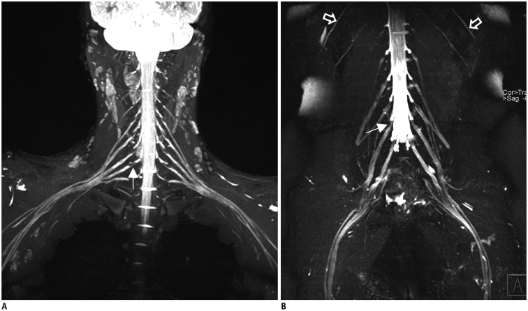
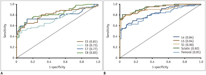
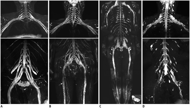
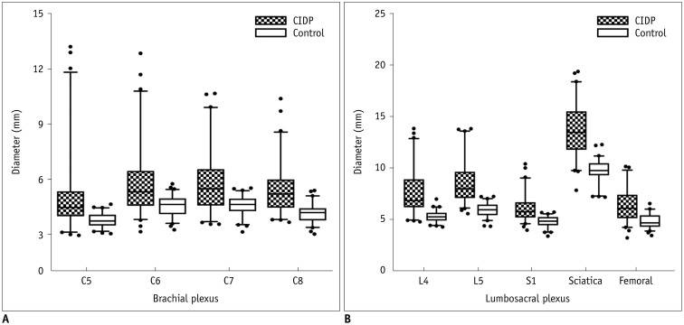
The bilateral extracranial vagus (n = 11), trigeminal (n = 12), and intercostal nerves (n = 10) were hypertrophic. Plexus hypertrophies were observed in the brachial plexus of 19 patients (61.3%) and in the LS plexus of 25 patients (80.6%). Patterns of hypertrophy included uniform hypertrophy (17 [54.8%] brachial plexuses and 21 [67.7%] LS plexuses), and multifocal fusiform hypertrophy (2 [6.5%] brachial plexuses and 4 [12.9%] LS plexuses) was present. Enlarged and/or contrast-enhanced cauda equina was found in 3 (9.7%) and 13 (41.9%) patients, respectively. Diameters of the brachial and LS nerve roots were significantly larger in CIDP than in controls ( < 0.001). The largest AUC was obtained for the L5 nerve. There were no significant differences in the course duration, I-RODS score, or diameter between patients with and without hypertrophy.

CONCLUSION

MRN is useful for the assessment of distribution and characteristics of the peripheral nerves in CIDP. Compared to other regions, LS plexus neurography is more sensitive for CIDP.

摘要

目的

使用磁共振神经成像(MRN)评估慢性炎症性脱髓鞘性多发性神经病(CIDP)中周围神经异常的分布和特征,并检验其诊断效率。

材料与方法

31 例 CIDP 患者和 21 例对照者接受了 MR 扫描。分别对臂丛和腰骶丛(LS)以及马尾进行三维采样完美,应用不同翻转角演化和 T1-/T2-加权涡轮自旋回波序列进行应用优化对比的神经成像。获得 CIDP 患者的临床数据和炎症 Rasch 构建整体残疾评分(I-RODS)评分。

结果

11 例双侧颅外迷走神经(n = 11)、12 例三叉神经(n = 12)和 10 例肋间神经(n = 10)呈肥大性改变。19 例患者(61.3%)出现臂丛神经丛肥大,25 例患者(80.6%)出现 LS 神经丛肥大。肥大模式包括均匀性肥大(17 个臂丛和 21 个 LS 神经丛,54.8%)和多灶性梭形肥大(2 个臂丛和 4 个 LS 神经丛,6.5%)。3 例(9.7%)和 13 例(41.9%)患者分别发现增大和/或对比增强的马尾。CIDP 患者的臂丛和 LS 神经根直径明显大于对照组(<0.001)。L5 神经根的 AUC 最大。有肥大和无肥大患者的病程持续时间、I-RODS 评分或直径无显著差异。

结论

MRN 可用于评估 CIDP 中周围神经的分布和特征。与其他区域相比,LS 神经丛神经成像对 CIDP 更敏感。

https://cdn.ncbi.nlm.nih.gov/pmc/blobs/e50a/7082655/35d56e7f0a0f/kjr-21-483-g005.jpg
https://cdn.ncbi.nlm.nih.gov/pmc/blobs/e50a/7082655/cee291b7bd8c/kjr-21-483-g001.jpg
https://cdn.ncbi.nlm.nih.gov/pmc/blobs/e50a/7082655/cd0af540c904/kjr-21-483-g002.jpg
https://cdn.ncbi.nlm.nih.gov/pmc/blobs/e50a/7082655/d528dfc3b9eb/kjr-21-483-g003.jpg
https://cdn.ncbi.nlm.nih.gov/pmc/blobs/e50a/7082655/cdf95a72e9d9/kjr-21-483-g004.jpg
https://cdn.ncbi.nlm.nih.gov/pmc/blobs/e50a/7082655/35d56e7f0a0f/kjr-21-483-g005.jpg

相似文献

1
Use of Magnetic Resonance Neurography for Evaluating the Distribution and Patterns of Chronic Inflammatory Demyelinating Polyneuropathy.应用磁共振神经成像评估慢性炎症性脱髓鞘性多发性神经病的分布和类型。
Korean J Radiol. 2020 Apr;21(4):483-493. doi: 10.3348/kjr.2019.0739.
2
Multisequence Quantitative Magnetic Resonance Neurography of Brachial and Lumbosacral Plexus in Chronic Inflammatory Demyelinating Polyneuropathy.慢性炎症性脱髓鞘性多发性神经病中臂丛和腰骶丛的多序列定量磁共振神经成像
Front Neurosci. 2021 Jul 23;15:649071. doi: 10.3389/fnins.2021.649071. eCollection 2021.
3
The Noninvasive Diagnostic Value of MRN for CIDP: A Research from Qualitative to Quantitative.磁共振神经成像技术(MRN)对 CIDP 的无创诊断价值:从定性到定量的研究。
Spine (Phila Pa 1976). 2020 Nov 1;45(21):1506-1512. doi: 10.1097/BRS.0000000000003599.
4
Simultaneous MR neurography and apparent T2 mapping in brachial plexus: Evaluation of patients with chronic inflammatory demyelinating polyradiculoneuropathy.臂丛神经的同步磁共振神经成像和表观T2映射:慢性炎性脱髓鞘性多发性神经根神经病患者的评估
Magn Reson Imaging. 2019 Jan;55:112-117. doi: 10.1016/j.mri.2018.09.025. Epub 2018 Sep 25.
5
Magnetic resonance neurography in diagnosing childhood chronic inflammatory demyelinating polyradiculoneuropathy.磁共振神经成像在儿童慢性炎症性脱髓鞘性多发神经根神经病诊断中的应用。
Brain Dev. 2021 Feb;43(2):352-356. doi: 10.1016/j.braindev.2020.10.001. Epub 2020 Oct 21.
6
Large coverage MR neurography in CIDP: diagnostic accuracy and electrophysiological correlation.慢性炎性脱髓鞘性多发性神经根神经病的大范围磁共振神经成像:诊断准确性及电生理相关性
J Neurol. 2017 Jul;264(7):1434-1443. doi: 10.1007/s00415-017-8543-7. Epub 2017 Jun 15.
7
MR neurography of lumbosacral nerve roots: Diagnostic value in chronic inflammatory demyelinating polyradiculoneuropathy and correlation with electrophysiological parameters.腰骶神经根磁共振神经成像:在慢性炎症性脱髓鞘性多发性神经根神经病中的诊断价值及与电生理参数的相关性。
Eur J Radiol. 2020 Mar;124:108816. doi: 10.1016/j.ejrad.2020.108816. Epub 2020 Jan 3.
8
MR neurography for the evaluation of CIDP.用于评估慢性炎性脱髓鞘性多发性神经病的磁共振神经成像
Muscle Nerve. 2017 Apr;55(4):483-489. doi: 10.1002/mus.25368. Epub 2016 Dec 26.
9
Prospective study of the additional benefit of plexus magnetic resonance imaging in the diagnosis of chronic inflammatory demyelinating polyneuropathy.前瞻性研究丛磁共振成像在慢性炎症性脱髓鞘性多发性神经病诊断中的附加益处。
Eur J Neurol. 2020 Jan;27(1):181-187. doi: 10.1111/ene.14053. Epub 2019 Aug 12.
10
Whole-body magnetic resonance neurography in patients with chronic inflammatory demyelinating polyneuropathy.慢性炎症性脱髓鞘性多发性神经病患者的全身磁共振神经成像。
Muscle Nerve. 2024 Jul;70(1):101-110. doi: 10.1002/mus.28098. Epub 2024 May 3.

引用本文的文献

1
Improved visualization of median, ulnar nerves, and small branches in the wrist and palm using contrast-enhanced magnetic resonance neurography.使用对比增强磁共振神经成像改善腕部和手掌正中神经、尺神经及小分支的可视化。
Ther Adv Neurol Disord. 2024 Mar 26;17:17562864241239739. doi: 10.1177/17562864241239739. eCollection 2024.
2
The diagnostic value of quantitative assessment of MR neurography in chronic inflammatory demyelinating polyradiculoneuropathy: a systematic review and meta-analysis.磁共振神经成像定量评估在慢性炎症性脱髓鞘性多发性神经根神经病中的诊断价值:系统评价和荟萃分析。
Br J Radiol. 2023 Nov;96(1151):20221037. doi: 10.1259/bjr.20221037. Epub 2023 Jul 4.
3

本文引用的文献

1
Dentate Nucleus Signal Intensity Decrease on T1-weighted MR Images after Switching from Gadopentetate Dimeglumine to Gadobutrol.钆喷酸葡胺切换为钆布醇后 T1 加权磁共振图像齿状核信号强度降低。
Radiology. 2018 Jun;287(3):816-823. doi: 10.1148/radiol.2018171398. Epub 2018 Mar 14.
2
Nerve Ultrasound Predicts Treatment Response in Chronic Inflammatory Demyelinating Polyradiculoneuropathy-a Prospective Follow-Up.神经超声预测慢性炎症性脱髓鞘性多发性神经病的治疗反应:一项前瞻性随访研究。
Neurotherapeutics. 2018 Apr;15(2):439-451. doi: 10.1007/s13311-018-0609-4.
3
CIDP and other inflammatory neuropathies in diabetes - diagnosis and management.
Role of MR Neurography for Evaluation of the Lumbosacral Plexus: A Scoping Review.
磁共振神经成像在腰骶丛评估中的作用:一项范围综述
J Korean Soc Radiol. 2022 Nov;83(6):1273-1285. doi: 10.3348/jksr.2022.0001. Epub 2022 Nov 15.
4
Infiltration of the spinal cord and peripheral nerves in multiple myeloma.多发性骨髓瘤中的脊髓和周围神经浸润。
Front Oncol. 2022 Oct 6;12:991246. doi: 10.3389/fonc.2022.991246. eCollection 2022.
5
Magnetic resonance tractography of the brachial plexus: step-by-step.臂丛神经的磁共振神经成像:分步指南
Quant Imaging Med Surg. 2022 Sep;12(9):4488-4501. doi: 10.21037/qims-22-30.
6
A Rare Phenotype of Uncommon Charcot-Marie-Tooth Genotypes Complicated With Inflammation Evaluated by Genetics and Magnetic Resonance Neurography.一种罕见表型的不常见遗传性运动感觉神经病基因型合并炎症,通过遗传学和磁共振神经造影术评估。
Front Genet. 2022 Jul 7;13:873641. doi: 10.3389/fgene.2022.873641. eCollection 2022.
7
A rare presentation of POEMS syndrome on magnetic resonance neurography: a case series.磁共振神经成像上POEMS综合征的罕见表现:病例系列
Ther Adv Neurol Disord. 2022 Jun 28;15:17562864221106350. doi: 10.1177/17562864221106350. eCollection 2022.
8
Chronic inflammatory demyelinating polyneuropathy: A unique case of chronic disease with atypical features.慢性炎症性脱髓鞘性多发性神经病:一例具有非典型特征的独特慢性病病例。
Radiol Case Rep. 2022 May 9;17(7):2441-2447. doi: 10.1016/j.radcr.2022.03.029. eCollection 2022 Jul.
9
Magnetic resonance tractography of the lumbosacral plexus: Step-by-step.腰骶丛磁共振神经束成像:分步解读。
Medicine (Baltimore). 2021 Feb 12;100(6):e24646. doi: 10.1097/MD.0000000000024646.
糖尿病相关 CIDP 和其他炎性神经病——诊断与管理。
Nat Rev Neurol. 2017 Oct;13(10):599-611. doi: 10.1038/nrneurol.2017.123. Epub 2017 Sep 15.
4
Brachial plexus magnetic resonance imaging differentiates between inflammatory neuropathies and does not predict disease course.臂丛磁共振成像可区分炎症性神经病,且不能预测疾病进程。
Brain Behav. 2017 Apr 4;7(5):e00632. doi: 10.1002/brb3.632. eCollection 2017 May.
5
High-resolution Imaging of Neural Anatomy and Pathology of the Neck.颈部神经解剖与病理学的高分辨率成像
Korean J Radiol. 2017 Jan-Feb;18(1):180-193. doi: 10.3348/kjr.2017.18.1.180. Epub 2017 Jan 5.
6
Clinical characteristics, and impairment and disability scale scores for different CIDP Disease Activity Status classes.不同慢性炎性脱髓鞘性多发性神经病(CIDP)疾病活动状态类别的临床特征、损伤和残疾量表评分
J Neurol Sci. 2017 Jan 15;372:223-227. doi: 10.1016/j.jns.2016.11.056. Epub 2016 Nov 23.
7
Evaluation of chronic inflammatory demyelinating polyneuropathy: 3D nerve-sheath signal increased with inked rest-tissue rapid acquisition of relaxation enhancement imaging (3D SHINKEI).慢性炎症性脱髓鞘性多发性神经病的评估:3D 神经鞘信号增加与墨迹静息组织弛豫增强成像(3D SHINKEI)。
Eur Radiol. 2017 Feb;27(2):447-453. doi: 10.1007/s00330-016-4406-3. Epub 2016 May 21.
8
The application of paramagnetic contrast-based T2 effect to 3D heavily T2W high-resolution MR imaging of the brachial plexus and its branches.基于顺磁性对比剂的T2效应在臂丛神经及其分支的三维重度T2加权高分辨率磁共振成像中的应用。
Eur J Radiol. 2016 Mar;85(3):578-84. doi: 10.1016/j.ejrad.2015.12.001. Epub 2015 Dec 4.
9
Chronic inflammatory demyelinating polyradiculoneuropathy: from pathology to phenotype.慢性炎症性脱髓鞘性多发性神经根神经病:从病理到表型
J Neurol Neurosurg Psychiatry. 2015 Sep;86(9):973-85. doi: 10.1136/jnnp-2014-309697. Epub 2015 Feb 12.
10
Diffusion-weighted MR neurography of the brachial and lumbosacral plexus: 3.0 T versus 1.5 T imaging.臂丛和腰骶丛的扩散加权磁共振神经成像:3.0T与1.5T成像对比
Eur J Radiol. 2015 Apr;84(4):696-702. doi: 10.1016/j.ejrad.2015.01.008. Epub 2015 Jan 17.