Elje Elisabeth, Mariussen Espen, Moriones Oscar H, Bastús Neus G, Puntes Victor, Kohl Yvonne, Dusinska Maria, Rundén-Pran Elise
Health Effects Laboratory, Department for Environmental Chemistry, NILU-Norwegian Institute for Air Research, Instituttveien 18, 2007 Kjeller, Norway.
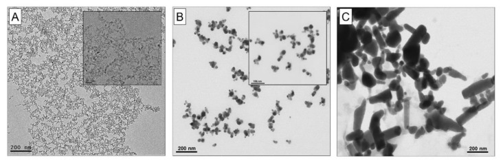
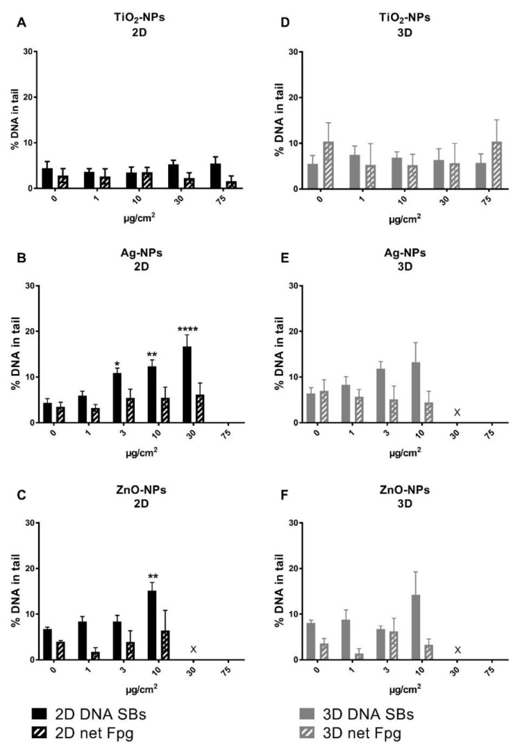
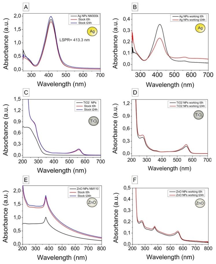
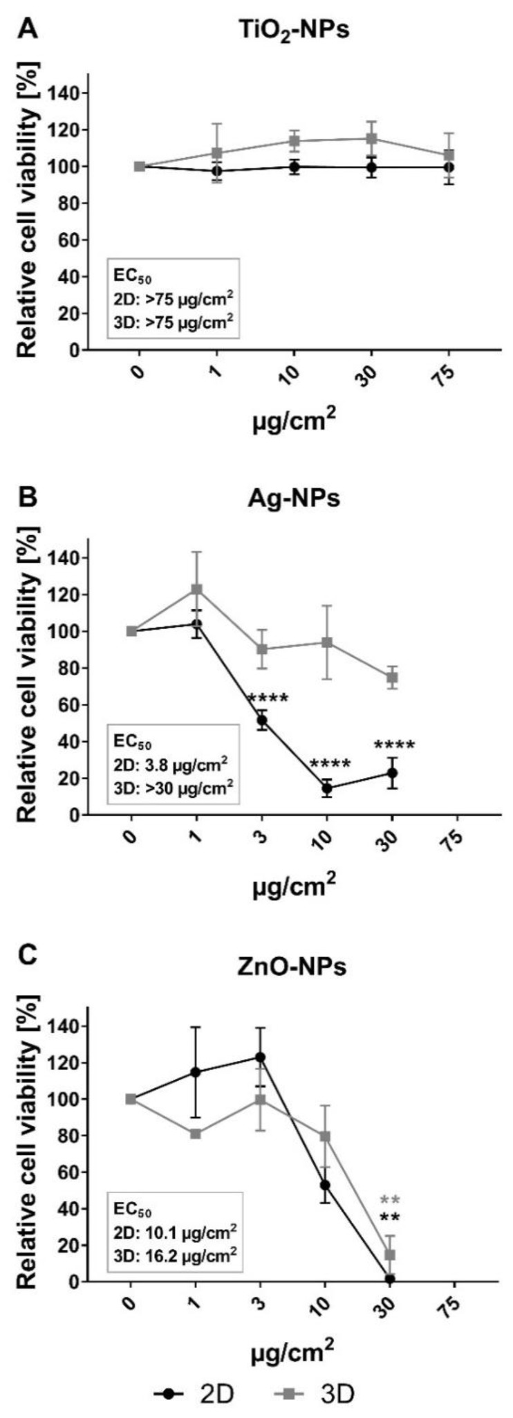
Department of Molecular Medicine, Institute of Basic Medical Sciences, Faculty of Medicine, University of Oslo, Sognsvannsveien 9, 0372 Oslo, Norway.
Nanomaterials (Basel). 2020 Mar 18;10(3):545. doi: 10.3390/nano10030545.
(1) In compliance with the 3Rs policy to reduce, refine and replace animal experiments, the development of advanced in vitro models is needed for nanotoxicity assessment. Cells cultivated in 3D resemble organ structures better than 2D cultures. This study aims to compare cytotoxic and genotoxic responses induced by titanium dioxide (TiO), silver (Ag) and zinc oxide (ZnO) nanoparticles (NPs) in 2D monolayer and 3D spheroid cultures of HepG2 human liver cells. (2) NPs were characterized by electron microscopy, dynamic light scattering, laser Doppler anemometry, UV-vis spectroscopy and mass spectrometry. Cytotoxicity was investigated by the alamarBlue assay and confocal microscopy in HepG2 monolayer and spheroid cultures after 24 h of NP exposure. DNA damage (strand breaks and oxidized base lesions) was measured by the comet assay. (3) Ag-NPs were aggregated at 24 h, and a substantial part of the ZnO-NPs was dissolved in culture medium. Ag-NPs induced stronger cytotoxicity in 2D cultures (EC 3.8 µg/cm) than in 3D cultures (EC > 30 µg/cm), and ZnO-NPs induced cytotoxicity to a similar extent in both models (EC 10.1-16.2 µg/cm). Ag- and ZnO-NPs showed a concentration-dependent genotoxic effect, but the effect was not statistically significant. TiO-NPs showed no toxicity (EC > 75 µg/cm). (4) This study shows that the HepG2 spheroid model is a promising advanced in vitro model for toxicity assessment of NPs.
(1) 为了遵守减少、优化和替代动物实验的3R政策,需要开发先进的体外模型用于纳米毒性评估。与二维培养相比,在三维环境中培养的细胞更类似于器官结构。本研究旨在比较二氧化钛(TiO)、银(Ag)和氧化锌(ZnO)纳米颗粒(NPs)在HepG2人肝细胞的二维单层和三维球体培养中诱导的细胞毒性和基因毒性反应。(2) 通过电子显微镜、动态光散射、激光多普勒测速仪、紫外可见光谱和质谱对纳米颗粒进行表征。在纳米颗粒暴露24小时后,通过alamarBlue测定法和共聚焦显微镜研究HepG2单层和球体培养中的细胞毒性。通过彗星试验测量DNA损伤(链断裂和氧化碱基损伤)。(3) 银纳米颗粒在24小时时发生聚集,并且相当一部分氧化锌纳米颗粒溶解在培养基中。银纳米颗粒在二维培养(EC 3.8 µg/cm)中比在三维培养(EC > 30 µg/cm)中诱导更强的细胞毒性,并且氧化锌纳米颗粒在两种模型中诱导的细胞毒性程度相似(EC 10.1 - 16.2 µg/cm)。银和氧化锌纳米颗粒显示出浓度依赖性的基因毒性作用,但该作用无统计学意义。二氧化钛纳米颗粒无毒性(EC > 75 µg/cm)。(4) 本研究表明,HepG2球体模型是一种有前景的先进体外模型,用于纳米颗粒的毒性评估。