• 文献检索
  • 文档翻译
  • 深度研究
  • 学术资讯
  • Suppr Zotero 插件Zotero 插件
  • 邀请有礼
  • 套餐&价格
  • 历史记录
应用&插件
Suppr Zotero 插件Zotero 插件浏览器插件Mac 客户端Windows 客户端微信小程序
定价
高级版会员购买积分包购买API积分包
服务
文献检索文档翻译深度研究API 文档MCP 服务
关于我们
关于 Suppr公司介绍联系我们用户协议隐私条款
关注我们

Suppr 超能文献

核心技术专利:CN118964589B侵权必究
粤ICP备2023148730 号-1Suppr @ 2026

文献检索

告别复杂PubMed语法,用中文像聊天一样搜索,搜遍4000万医学文献。AI智能推荐,让科研检索更轻松。

立即免费搜索

文件翻译

保留排版,准确专业,支持PDF/Word/PPT等文件格式,支持 12+语言互译。

免费翻译文档

深度研究

AI帮你快速写综述,25分钟生成高质量综述,智能提取关键信息,辅助科研写作。

立即免费体验

通过层层法和 3D 打印机构建具有定向的血管化心脏组织。

Vascularized cardiac tissue construction with orientation by layer-by-layer method and 3D printer.

机构信息

Building Block Science Joint Research Chair, Graduate School of Frontier Biosciences, Osaka University, 1-3 Yamadaoka, Suita, 565-0871, Japan.

出版信息

Sci Rep. 2020 Mar 26;10(1):5484. doi: 10.1038/s41598-020-59371-y.

DOI:10.1038/s41598-020-59371-y
PMID:32218447
原文链接:https://pmc.ncbi.nlm.nih.gov/articles/PMC7098983/
Abstract

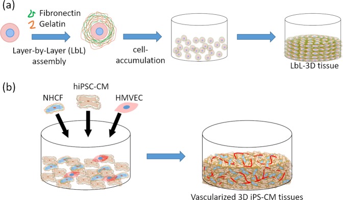
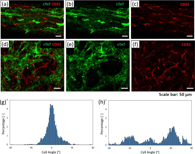
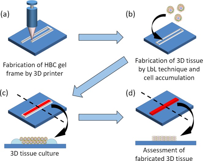
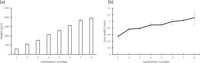
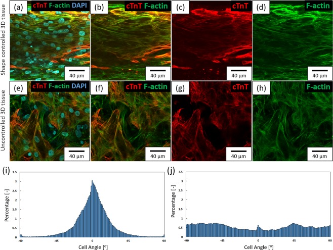
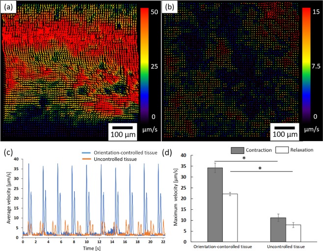
Herein, we report the fabrication of native organ-like three-dimensional (3D) cardiac tissue with an oriented structure and vascular network using a layer-by-layer (LbL), cell accumulation and 3D printing technique for regenerative medicine and pharmaceutical applications. We firstly evaluated the 3D shaping ability of hydroxybutyl chitosan (HBC), a thermoresponsive polymer, by using a robotic dispensing 3D printer. Next, we tried to fabricate orientation-controlled 3D cardiac tissue using human induced pluripotent stem cell-derived cardiomyocytes (hiPSC-CM) and normal human cardiac fibroblasts (NHCF) coated with extracellular matrix (ECM) nanofilms by layer-by-layer technique. These cells were seeded in the fabricated rectangular shape HBC gel frame. After cultivation of the fabricated tissue, fluorescence staining of the cytoskeleton revealed that hiPSC-CM and NHCF were aligned in one direction. Moreover, we were able to measure its contractile behavior using a video image analysis system. These results indicate that orientation-controlled cardiac tissue has more remarkable contractile function than uncontrolled cardiac tissue. Finally, co-culture with human cardiac microvascular endothelial cells (HMVEC) successfully provided a vascular network in orientation-controlled 3D cardiac tissue. The constructed 3D cardiac tissue with an oriented structure and vascular network would be a useful tool for regenerative medicine and pharmaceutical applications.

摘要

在这里,我们报告了使用层层(LbL)、细胞积累和 3D 打印技术制造具有定向结构和血管网络的天然器官样三维(3D)心脏组织的方法,用于再生医学和药物应用。我们首先使用机器人分配 3D 打印机评估了热响应聚合物羟丁基壳聚糖(HBC)的 3D 成型能力。接下来,我们尝试使用细胞外基质(ECM)纳米膜包被的人诱导多能干细胞衍生的心肌细胞(hiPSC-CM)和正常人心肌成纤维细胞(NHCF)通过层层技术制造定向控制的 3D 心脏组织。这些细胞被接种在制造的矩形 HBC 凝胶框架中。在制造的组织培养后,细胞骨架的荧光染色显示 hiPSC-CM 和 NHCF 沿一个方向排列。此外,我们能够使用视频图像分析系统测量其收缩行为。这些结果表明,定向控制的心脏组织比非定向控制的心脏组织具有更显著的收缩功能。最后,与人心脏微血管内皮细胞(HMVEC)的共培养成功地在定向控制的 3D 心脏组织中提供了血管网络。具有定向结构和血管网络的构建 3D 心脏组织将成为再生医学和药物应用的有用工具。

https://cdn.ncbi.nlm.nih.gov/pmc/blobs/4d79/7098983/2ea5c56eff20/41598_2020_59371_Fig6_HTML.jpg
https://cdn.ncbi.nlm.nih.gov/pmc/blobs/4d79/7098983/2d8975680b70/41598_2020_59371_Fig1_HTML.jpg
https://cdn.ncbi.nlm.nih.gov/pmc/blobs/4d79/7098983/ff746d1cd477/41598_2020_59371_Fig2_HTML.jpg
https://cdn.ncbi.nlm.nih.gov/pmc/blobs/4d79/7098983/e7f165ad87e8/41598_2020_59371_Fig3_HTML.jpg
https://cdn.ncbi.nlm.nih.gov/pmc/blobs/4d79/7098983/a0c51f085201/41598_2020_59371_Fig4_HTML.jpg
https://cdn.ncbi.nlm.nih.gov/pmc/blobs/4d79/7098983/e969f91806e1/41598_2020_59371_Fig5_HTML.jpg
https://cdn.ncbi.nlm.nih.gov/pmc/blobs/4d79/7098983/2ea5c56eff20/41598_2020_59371_Fig6_HTML.jpg

相似文献

1
Vascularized cardiac tissue construction with orientation by layer-by-layer method and 3D printer.通过层层法和 3D 打印机构建具有定向的血管化心脏组织。
Sci Rep. 2020 Mar 26;10(1):5484. doi: 10.1038/s41598-020-59371-y.
2
Fabrication of Orientation-Controlled 3D Tissues Using a Layer-by-Layer Technique and 3D Printed a Thermoresponsive Gel Frame.采用层层技术和 3D 打印的热响应凝胶框架制造具有定向控制的 3D 组织。
Tissue Eng Part C Methods. 2017 Jun;23(6):357-366. doi: 10.1089/ten.TEC.2017.0134. Epub 2017 Jun 5.
3
Development of vascularized iPSC derived 3D-cardiomyocyte tissues by filtration Layer-by-Layer technique and their application for pharmaceutical assays.通过逐层过滤技术构建血管化诱导多能干细胞衍生的3D心肌组织及其在药物检测中的应用。
Acta Biomater. 2016 Mar;33:110-21. doi: 10.1016/j.actbio.2016.01.033. Epub 2016 Jan 25.
4
Fabrication of Cardiac Constructs Using Bio-3D Printer.使用生物 3D 打印机制作心脏构建体。
Methods Mol Biol. 2021;2320:53-63. doi: 10.1007/978-1-0716-1484-6_6.
5
Supersensitive Layer-by-Layer 3D Cardiac Tissues Fabricated on a Collagen Culture Vessel Using Human-Induced Pluripotent Stem Cells.利用人诱导多能干细胞在胶原培养容器上构建超敏感的层状 3D 心脏组织。
Tissue Eng Part C Methods. 2020 Sep;26(9):493-502. doi: 10.1089/ten.TEC.2020.0195.
6
A multi-cellular 3D bioprinting approach for vascularized heart tissue engineering based on HUVECs and iPSC-derived cardiomyocytes.基于 HUVECs 和 iPSC 分化的心肌细胞的用于血管化心脏组织工程的多细胞 3D 生物打印方法。
Sci Rep. 2018 Sep 10;8(1):13532. doi: 10.1038/s41598-018-31848-x.
7
Myocardial Tissue Engineering With Cells Derived From Human-Induced Pluripotent Stem Cells and a Native-Like, High-Resolution, 3-Dimensionally Printed Scaffold.利用源自人诱导多能干细胞和类似天然的、高分辨率三维打印支架的细胞进行心肌组织工程
Circ Res. 2017 Apr 14;120(8):1318-1325. doi: 10.1161/CIRCRESAHA.116.310277. Epub 2017 Jan 9.
8
Engineered heart tissue models from hiPSC-derived cardiomyocytes and cardiac ECM for disease modeling and drug testing applications.基于人诱导多能干细胞(hiPSC)分化的心肌细胞和心脏细胞外基质构建的工程化心脏组织模型,可用于疾病建模和药物测试应用。
Acta Biomater. 2019 Jul 1;92:145-159. doi: 10.1016/j.actbio.2019.05.016. Epub 2019 May 7.
9
Functional 3-D cardiac co-culture model using bioactive chitosan nanofiber scaffolds.使用生物活性壳聚糖纳米纤维支架构建功能性 3D 心脏共培养模型。
Biotechnol Bioeng. 2013 Feb;110(2):637-47. doi: 10.1002/bit.24727. Epub 2012 Oct 5.
10
Bioprinting 3D microfibrous scaffolds for engineering endothelialized myocardium and heart-on-a-chip.用于构建内皮化心肌和芯片心脏的生物打印3D微纤维支架
Biomaterials. 2016 Dec;110:45-59. doi: 10.1016/j.biomaterials.2016.09.003. Epub 2016 Sep 5.

引用本文的文献

1
Harnessing native blueprints for designing bioinks to bioprint functional cardiac tissue.利用天然蓝图设计生物墨水以生物打印功能性心脏组织。
iScience. 2025 Jan 23;28(3):111882. doi: 10.1016/j.isci.2025.111882. eCollection 2025 Mar 21.
2
Advances and Prospects in Using Induced Pluripotent Stem Cells for 3D Bioprinting in Cardiac Tissue Engineering.诱导多能干细胞在心脏组织工程3D生物打印中的研究进展与展望
Rev Cardiovasc Med. 2025 Mar 19;26(3):26697. doi: 10.31083/RCM26697. eCollection 2025 Mar.
3
Layer-by-Layer Nanoarchitectonics: A Method for Everything in Layered Structures.

本文引用的文献

1
Porous, Ventricular Extracellular Matrix-Derived Foams as a Platform for Cardiac Cell Culture.多孔心室细胞外基质衍生泡沫作为心脏细胞培养平台
Biores Open Access. 2015 Oct 1;4(1):374-88. doi: 10.1089/biores.2015.0030. eCollection 2015.
2
Image-based evaluation of contraction-relaxation kinetics of human-induced pluripotent stem cell-derived cardiomyocytes: Correlation and complementarity with extracellular electrophysiology.基于图像的人诱导多能干细胞衍生心肌细胞收缩-松弛动力学评估:与细胞外电生理学的相关性和互补性。
J Mol Cell Cardiol. 2014 Dec;77:178-91. doi: 10.1016/j.yjmcc.2014.09.010. Epub 2014 Sep 23.
3
逐层纳米结构构建:一种用于层状结构中万物的方法。
Materials (Basel). 2025 Feb 1;18(3):654. doi: 10.3390/ma18030654.
4
Bioprinting approaches in cardiac tissue engineering to reproduce blood-pumping heart function.心脏组织工程中的生物打印方法,用于重现具有血液泵送功能的心脏。
iScience. 2024 Dec 20;28(1):111664. doi: 10.1016/j.isci.2024.111664. eCollection 2025 Jan 17.
5
Recent advances in layer-by-layer assembly scaffolds for co-delivery of bioactive molecules for bone regeneration: an updated review.近年来,用于骨再生的生物活性分子共递送的层层组装支架的研究进展:更新综述。
J Transl Med. 2024 Nov 5;22(1):1001. doi: 10.1186/s12967-024-05809-0.
6
3D-bioprinted cardiac tissues and their potential for disease modeling.3D生物打印心脏组织及其疾病建模潜力。
J 3D Print Med. 2023 Jun;7(2). doi: 10.2217/3dp-2022-0023. Epub 2023 Apr 4.
7
Construction of millimeter-scale vascularized engineered myocardial tissue using a mixed gel.使用混合凝胶构建毫米级血管化工程心肌组织。
Regen Biomater. 2023 Dec 29;11:rbad117. doi: 10.1093/rb/rbad117. eCollection 2024.
8
Heart-on-a-chip systems with tissue-specific functionalities for physiological, pathological, and pharmacological studies.具有组织特异性功能的芯片上心脏系统,用于生理、病理和药理研究。
Mater Today Bio. 2023 Dec 20;24:100914. doi: 10.1016/j.mtbio.2023.100914. eCollection 2024 Feb.
9
Recent Developments in Layer-by-Layer Assembly for Drug Delivery and Tissue Engineering Applications.层状组装技术在药物传递和组织工程应用中的最新进展。
Adv Healthc Mater. 2024 Mar;13(8):e2302713. doi: 10.1002/adhm.202302713. Epub 2024 Jan 7.
10
Generation of Patterned Cocultures in 2D and 3D: State of the Art.二维和三维模式共培养的生成:当前技术水平
ACS Omega. 2023 Sep 13;8(38):34249-34261. doi: 10.1021/acsomega.3c02713. eCollection 2023 Sep 26.
Excitation propagation in three-dimensional engineered hearts using decellularized extracellular matrix.
使用脱细胞细胞外基质的三维工程心脏中的兴奋传播。
Biomaterials. 2014 Sep;35(27):7839-50. doi: 10.1016/j.biomaterials.2014.05.080. Epub 2014 Jun 20.
4
Micropattern width dependent sarcomere development in human ESC-derived cardiomyocytes.人胚胎干细胞衍生心肌细胞中微图案宽度依赖的肌节发育
Biomaterials. 2014 May;35(15):4454-64. doi: 10.1016/j.biomaterials.2014.02.001. Epub 2014 Feb 28.
5
Young developmental age cardiac extracellular matrix promotes the expansion of neonatal cardiomyocytes in vitro.年轻发育期心脏细胞外基质促进体外新生大鼠心肌细胞的扩增。
Acta Biomater. 2014 Jan;10(1):194-204. doi: 10.1016/j.actbio.2013.08.037. Epub 2013 Sep 6.
6
Repopulation of decellularized mouse heart with human induced pluripotent stem cell-derived cardiovascular progenitor cells.用人类诱导多能干细胞衍生的心血管祖细胞重构成去细胞化的小鼠心脏。
Nat Commun. 2013;4:2307. doi: 10.1038/ncomms3307.
7
Experimental investigation of collagen waviness and orientation in the arterial adventitia using confocal laser scanning microscopy.应用共聚焦激光扫描显微镜研究动脉外膜中胶原的波纹和方向。
Biomech Model Mechanobiol. 2012 Mar;11(3-4):461-73. doi: 10.1007/s10237-011-0325-z. Epub 2011 Jul 10.
8
Mesoscopic hydrogel molding to control the 3D geometry of bioartificial muscle tissues.介观水凝胶成型以控制生物人工肌肉组织的三维几何形状。
Nat Protoc. 2009;4(10):1522-34. doi: 10.1038/nprot.2009.155. Epub 2009 Sep 24.
9
Effect of aging on elastin functionality in human cerebral arteries.衰老对人脑动脉中弹性蛋白功能的影响。
Stroke. 2009 Jul;40(7):2552-6. doi: 10.1161/STROKEAHA.108.528091. Epub 2009 May 28.
10
Production of arrays of cardiac and skeletal muscle myofibers by micropatterning techniques on a soft substrate.通过在柔软基底上采用微图案化技术制备心肌和骨骼肌肌纤维阵列。
Biomed Microdevices. 2009 Apr;11(2):389-400. doi: 10.1007/s10544-008-9245-9.