• 文献检索
  • 文档翻译
  • 深度研究
  • 学术资讯
  • Suppr Zotero 插件Zotero 插件
  • 邀请有礼
  • 套餐&价格
  • 历史记录
应用&插件
Suppr Zotero 插件Zotero 插件浏览器插件Mac 客户端Windows 客户端微信小程序
定价
高级版会员购买积分包购买API积分包
服务
文献检索文档翻译深度研究API 文档MCP 服务
关于我们
关于 Suppr公司介绍联系我们用户协议隐私条款
关注我们

Suppr 超能文献

核心技术专利:CN118964589B侵权必究
粤ICP备2023148730 号-1Suppr @ 2026

文献检索

告别复杂PubMed语法,用中文像聊天一样搜索,搜遍4000万医学文献。AI智能推荐,让科研检索更轻松。

立即免费搜索

文件翻译

保留排版,准确专业,支持PDF/Word/PPT等文件格式,支持 12+语言互译。

免费翻译文档

深度研究

AI帮你快速写综述,25分钟生成高质量综述,智能提取关键信息,辅助科研写作。

立即免费体验

新生儿获得性后颅窝异常的超声表现

Ultrasound of acquired posterior fossa abnormalities in the newborn.

机构信息

NICU, Fondazione IRCCS Ca' Granda Ospedale Maggiore Policlinico, Milan, Italy.

University of Milan, Department of Clinical Sciences and Community Health, Milan, Italy.

出版信息

Pediatr Res. 2020 Mar;87(Suppl 1):25-36. doi: 10.1038/s41390-020-0778-9.

DOI:10.1038/s41390-020-0778-9
PMID:32218537
原文链接:https://pmc.ncbi.nlm.nih.gov/articles/PMC7098891/
Abstract

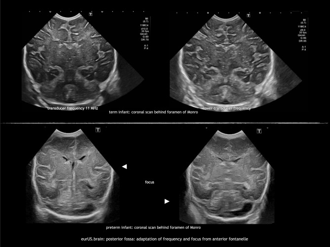
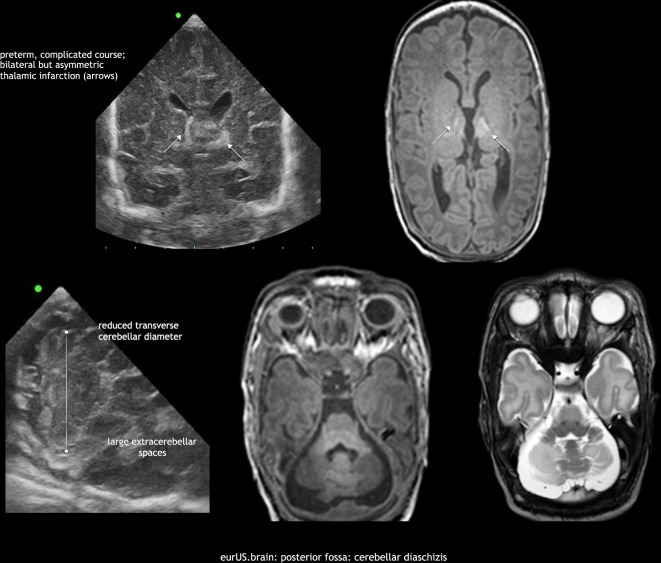
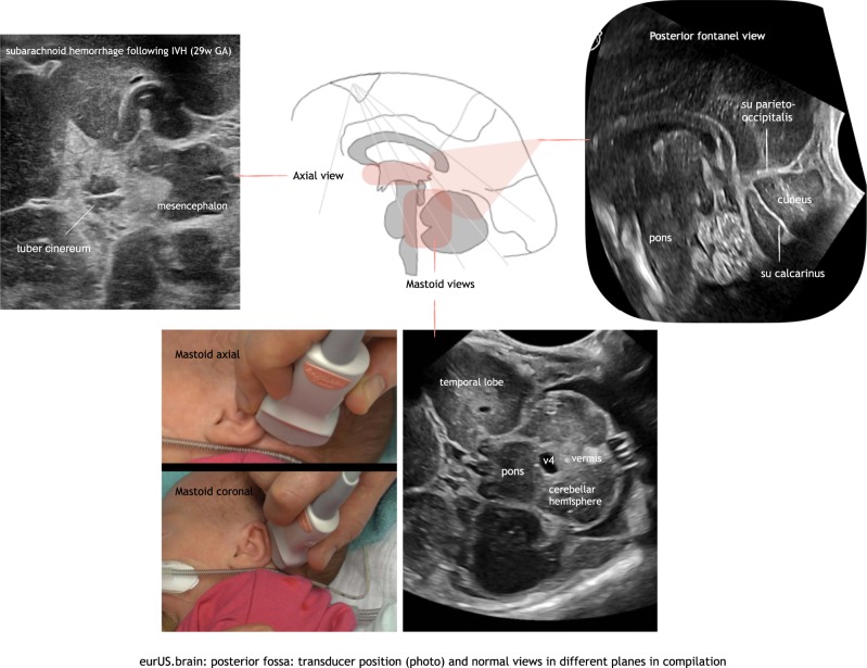
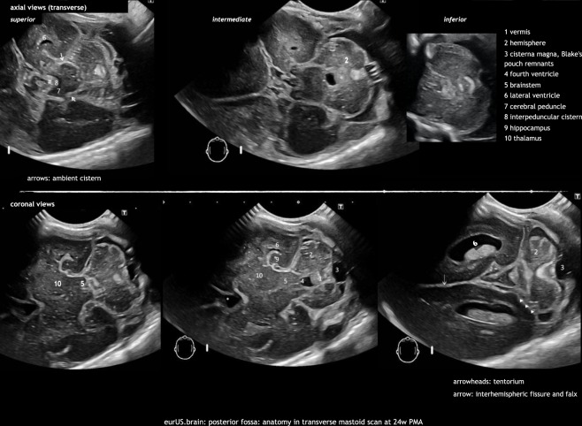
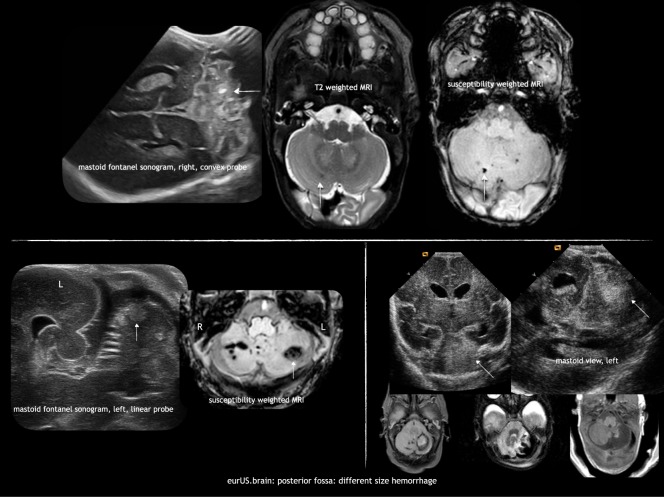
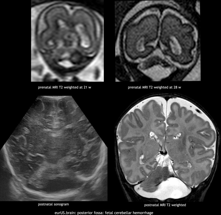
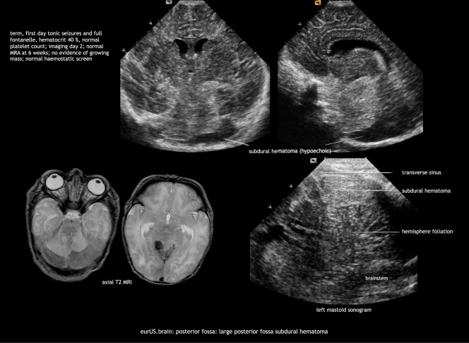
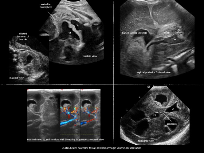
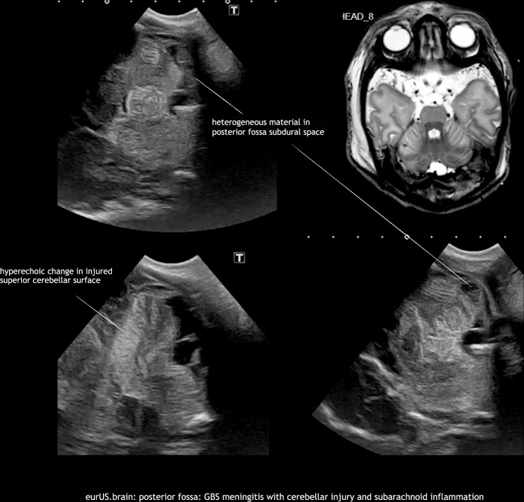
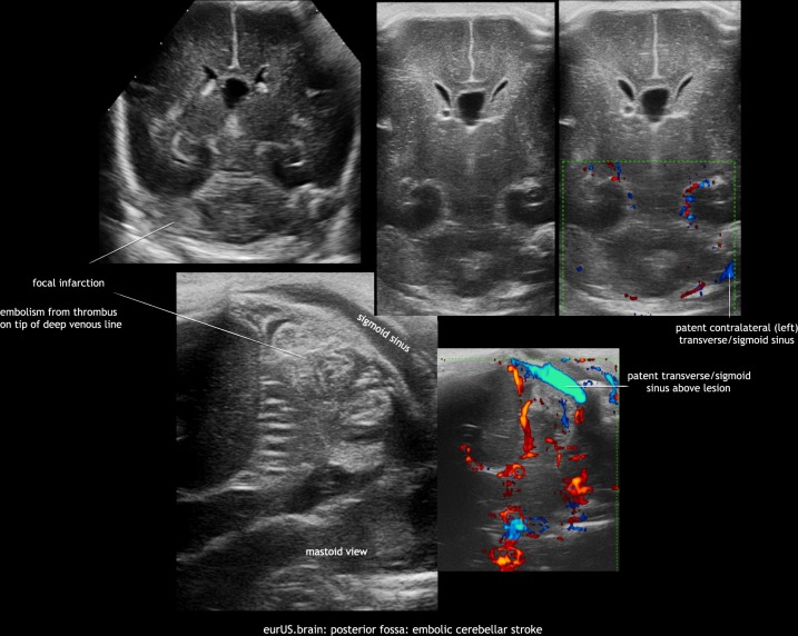
Neonatal brain sonography is part of routine clinical practice in neonatal intensive care units, but ultrasound imaging of the posterior fossa has gained increasing attention since the burden of perinatal acquired posterior fossa abnormalities and their impact on motor and cognitive neurodevelopmental outcome have been recognized. Although magnetic resonance imaging (MRI) is often superior, posterior fossa abnormalities can be suspected or detected by optimized cranial ultrasound (CUS) scans, which allow an early and bed-side diagnosis and monitoring through sequential scans over a long period of time. Different ultrasound appearances and injury patterns of posterior fossa abnormalities are described according to gestational age at birth and characteristics of the pathogenetic insult. The aim of this review article is to describe options to improve posterior fossa sequential CUS image quality, including the use of supplemental acoustic windows, to show standard views and normal ultrasound anatomy of the posterior fossa, and to describe the ultrasound characteristics of acquired posterior fossa lesions in preterm and term infants with effect on long-term outcome. The limitations and pitfalls of CUS and the role of MRI are discussed.

摘要

新生儿颅脑超声是新生儿重症监护病房常规临床实践的一部分,但由于认识到围产期获得性后颅窝异常及其对运动和认知神经发育结局的影响,后颅窝的超声成像越来越受到关注。虽然磁共振成像(MRI)通常更优越,但通过优化的头颅超声(CUS)扫描可以怀疑或检测到后颅窝异常,这允许通过在很长一段时间内进行连续扫描进行早期和床边诊断和监测。根据出生时的胎龄和致病因素的特点,描述了后颅窝异常的不同超声表现和损伤模式。本文的目的是描述改善后颅窝连续 CUS 图像质量的选择,包括使用补充的声学窗口,以显示后颅窝的标准视图和正常超声解剖结构,并描述早产儿和足月儿获得性后颅窝病变的超声特征及其对长期结局的影响。讨论了 CUS 的局限性和陷阱以及 MRI 的作用。

https://cdn.ncbi.nlm.nih.gov/pmc/blobs/8902/7098891/4640c4bcbeae/41390_2020_778_Fig11_HTML.jpg
https://cdn.ncbi.nlm.nih.gov/pmc/blobs/8902/7098891/5abfbe5e6ac6/41390_2020_778_Fig1_HTML.jpg
https://cdn.ncbi.nlm.nih.gov/pmc/blobs/8902/7098891/36b74659d1d0/41390_2020_778_Fig2_HTML.jpg
https://cdn.ncbi.nlm.nih.gov/pmc/blobs/8902/7098891/b9477d845bea/41390_2020_778_Fig3_HTML.jpg
https://cdn.ncbi.nlm.nih.gov/pmc/blobs/8902/7098891/f19ca75efd71/41390_2020_778_Fig4_HTML.jpg
https://cdn.ncbi.nlm.nih.gov/pmc/blobs/8902/7098891/9881480d8aab/41390_2020_778_Fig5_HTML.jpg
https://cdn.ncbi.nlm.nih.gov/pmc/blobs/8902/7098891/4c092e2bc425/41390_2020_778_Fig6_HTML.jpg
https://cdn.ncbi.nlm.nih.gov/pmc/blobs/8902/7098891/831d0509806b/41390_2020_778_Fig7_HTML.jpg
https://cdn.ncbi.nlm.nih.gov/pmc/blobs/8902/7098891/d2390f43590b/41390_2020_778_Fig8_HTML.jpg
https://cdn.ncbi.nlm.nih.gov/pmc/blobs/8902/7098891/95226ea72463/41390_2020_778_Fig9_HTML.jpg
https://cdn.ncbi.nlm.nih.gov/pmc/blobs/8902/7098891/876d65fe6def/41390_2020_778_Fig10_HTML.jpg
https://cdn.ncbi.nlm.nih.gov/pmc/blobs/8902/7098891/4640c4bcbeae/41390_2020_778_Fig11_HTML.jpg

相似文献

1
Ultrasound of acquired posterior fossa abnormalities in the newborn.新生儿获得性后颅窝异常的超声表现
Pediatr Res. 2020 Mar;87(Suppl 1):25-36. doi: 10.1038/s41390-020-0778-9.
2
A new pattern of cerebellar hemorrhages in preterm infants.早产儿小脑出血的一种新模式。
Pediatrics. 1998 Dec;102(6):E62. doi: 10.1542/peds.102.6.e62.
3
Cranial ultrasonography of the immature cerebellum: Role and limitations.未成熟小脑的头颅超声检查:作用与局限性
Semin Fetal Neonatal Med. 2016 Oct;21(5):295-304. doi: 10.1016/j.siny.2016.04.011. Epub 2016 May 14.
4
Posterior fossa abnormalities in high-risk term infants: comparison of ultrasound and MRI.高危足月儿的后颅窝异常:超声与磁共振成像的比较
Eur Radiol. 2015 Sep;25(9):2575-83. doi: 10.1007/s00330-015-3665-8. Epub 2015 Apr 22.
5
[Cranial ultrasound through posterolateral fontanelle in visualization of posterior fossa abnormalities in preterm and term neonates].[经后外侧囟门的颅脑超声检查用于观察早产和足月新生儿后颅窝异常情况]
Ginekol Pol. 2015 Oct;86(10):774-81. doi: 10.17772/gp/57860.
6
Ultrasound detection of posterior fossa abnormalities in full-term neonates.足月新生儿后颅窝异常的超声检测。
Early Hum Dev. 2012 Apr;88(4):233-9. doi: 10.1016/j.earlhumdev.2011.08.011. Epub 2011 Sep 15.
7
Diagnostic imaging of posterior fossa anomalies in the fetus.胎儿后颅窝异常的诊断性影像学检查
Semin Fetal Neonatal Med. 2016 Oct;21(5):312-20. doi: 10.1016/j.siny.2016.04.007. Epub 2016 May 18.
8
Cranial ultrasound findings in preterm germinal matrix haemorrhage, sequelae and outcome.早产儿脑室内出血、后遗症和结局的头颅超声表现。
Pediatr Res. 2020 Mar;87(Suppl 1):13-24. doi: 10.1038/s41390-020-0780-2.
9
Comparison of cranial ultrasound and MRI for detecting BRAIN injury in extremely preterm infants and correlation with neurological outcomes at 1 and 3 years.比较经颅超声与 MRI 对极早产儿脑损伤的检测及其与 1 岁及 3 岁时神经发育结局的相关性。
Eur J Pediatr. 2019 Jul;178(7):1053-1061. doi: 10.1007/s00431-019-03388-7. Epub 2019 May 7.
10
Risk factors and ultrasonographic profile of posterior fossa haemorrhages in preterm infants.早产儿后颅窝出血的危险因素及超声特征
J Paediatr Child Health. 2009 Apr;45(4):215-8. doi: 10.1111/j.1440-1754.2008.01456.x. Epub 2009 Mar 23.

引用本文的文献

1
Predictive model of ibuprofen treatment failure in very preterm infants with patent ductus arteriosus using machine learning techniques.运用机器学习技术建立极早产儿动脉导管未闭布洛芬治疗失败的预测模型。
J Perinatol. 2025 Jul 8. doi: 10.1038/s41372-025-02346-6.
2
Predictive value of transcranial Doppler ultrasound for brain development and craniocerebral injury in premature infants.经颅多普勒超声对早产儿脑发育及颅脑损伤的预测价值
Am J Transl Res. 2025 Mar 15;17(3):2076-2082. doi: 10.62347/QJZN6645. eCollection 2025.
3
Point-of-care brain ultrasound and transcranial doppler or color-coded doppler in critically ill neonates and children.

本文引用的文献

1
Involvement of the anterior lobe of the cerebellar vermis in perinatal profound hypoxia.小脑蚓部前叶在围产期严重缺氧中的受累情况。
AJNR Am J Neuroradiol. 2007 Jan;28(1):16-9.
2
Cerebellar hemorrhage in the preterm infant: ultrasonographic findings and risk factors.早产儿小脑出血:超声检查结果及危险因素
Pediatrics. 2005 Sep;116(3):717-24. doi: 10.1542/peds.2005-0556.
3
Posterior fontanelle sonography: an acoustic window into the neonatal brain.后囟超声检查:新生儿脑部的声学窗口
床边脑超声和经颅多普勒或彩色编码多普勒在危重新生儿和儿童中的应用。
Eur J Pediatr. 2024 Mar;183(3):1059-1072. doi: 10.1007/s00431-023-05388-0. Epub 2023 Dec 19.
4
An Overview of Cytomegalovirus Infection in Pregnancy.妊娠期巨细胞病毒感染概述
Diagnostics (Basel). 2022 Oct 7;12(10):2429. doi: 10.3390/diagnostics12102429.
5
Randomized Trial on Echocardiography-Guided Ductus Arteriosus Treatment to Reduce Necrotizing Enterocolitis.超声心动图引导下动脉导管治疗以降低坏死性小肠结肠炎的随机试验
Front Pediatr. 2022 Jan 28;9:807712. doi: 10.3389/fped.2021.807712. eCollection 2021.
6
Methods for Monitoring Risk of Hypoxic Damage in Fetal and Neonatal Brains: A Review.监测胎儿和新生儿大脑缺氧损伤风险的方法:综述。
Fetal Diagn Ther. 2022;49(1-2):1-24. doi: 10.1159/000520987. Epub 2021 Dec 6.
7
Consensus Approach for Standardizing the Screening and Classification of Preterm Brain Injury Diagnosed With Cranial Ultrasound: A Canadian Perspective.颅超声诊断早产脑损伤的筛查与分类标准化的共识方法:加拿大视角
Front Pediatr. 2021 Mar 8;9:618236. doi: 10.3389/fped.2021.618236. eCollection 2021.
8
Cranial ultrasound by neonatologists.新生儿科医生进行的头颅超声检查。
Pediatr Res. 2020 Mar;87(Suppl 1):1-2. doi: 10.1038/s41390-020-0779-8.
AJNR Am J Neuroradiol. 2004 Aug;25(7):1274-82.
4
Severe fetal cytomegalovirus infection associated with cerebellar hemorrhage.严重胎儿巨细胞病毒感染伴小脑出血。
Ultrasound Obstet Gynecol. 2004 Apr;23(4):402-6. doi: 10.1002/uog.1021.
5
MR imaging quantification of cerebellar growth following hypoxic-ischemic injury to the neonatal brain.新生儿脑缺氧缺血性损伤后小脑生长的磁共振成像定量分析
AJNR Am J Neuroradiol. 2004 Mar;25(3):463-8.
6
Reproducibility and accuracy of MR imaging of the brain after severe birth asphyxia.重度出生窒息后脑磁共振成像的可重复性和准确性
AJNR Am J Neuroradiol. 1999 Aug;20(7):1343-8.
7
Personality characteristics and developmental history of creative college women.富有创造力的大学女生的个性特征与成长经历
Genet Psychol Monogr. 1967 Nov;76(2):205-56.
8
[Oral spirochetes antibodies observed in the human serum].[在人血清中观察到的口腔螺旋体抗体]
Shigaku. 1971 Feb;58(6):765-85.
9
[A curious trilogy: kidney cancer, polycystic kidney, and renal lithiasis].一个奇特的三联征:肾癌、多囊肾和肾结石
J Urol Nephrol (Paris). 1972 Jun;78(6):548-9.
10
Fatty acid composition of Spirochaeta stenostrepta.狭窄螺旋体的脂肪酸组成。
J Bacteriol. 1972 Oct;112(1):629-31. doi: 10.1128/jb.112.1.629-631.1972.