Mohammad Khorshid, Scott James N, Leijser Lara M, Zein Hussein, Afifi Jehier, Piedboeuf Bruno, de Vries Linda S, van Wezel-Meijler Gerda, Lee Shoo K, Shah Prakesh S
Department of Pediatrics, University of Calgary, Calgary, AB, Canada.
Departments of Diagnostic Imaging and Clinical Neurosciences, University of Calgary, Calgary, AB, Canada.
Front Pediatr. 2021 Mar 8;9:618236. doi: 10.3389/fped.2021.618236. eCollection 2021.
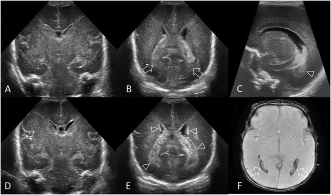
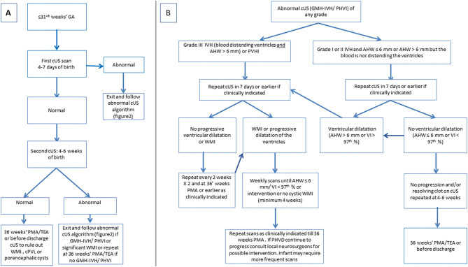
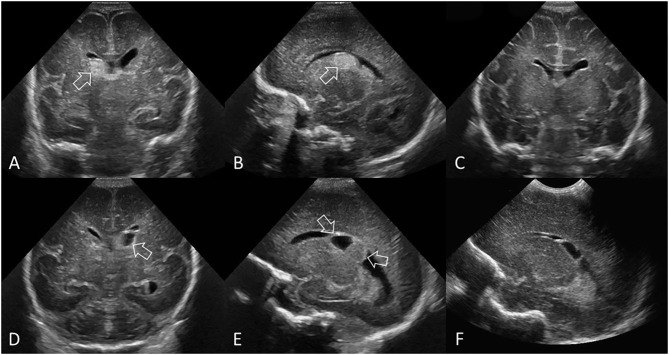
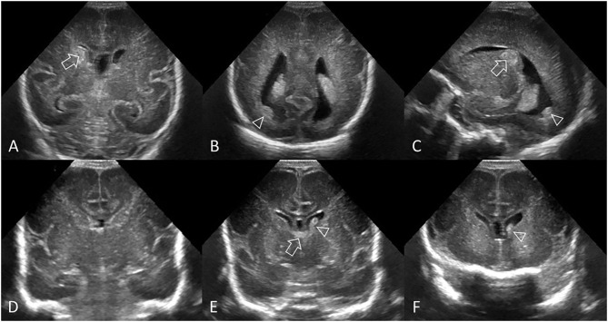
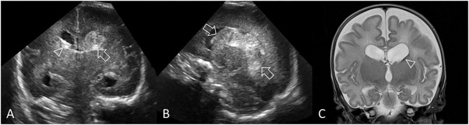
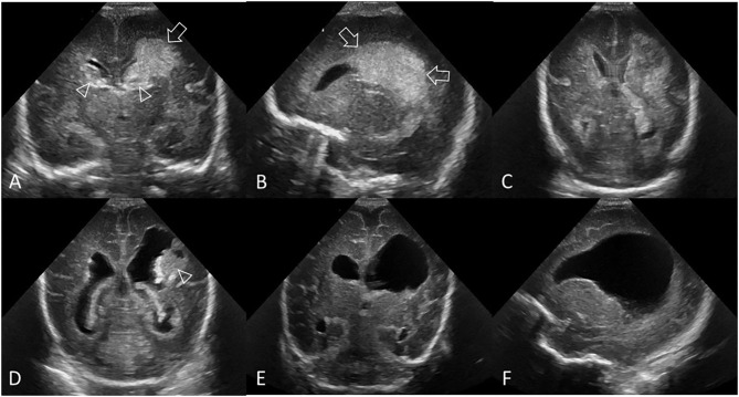
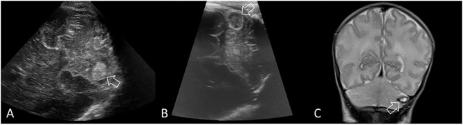
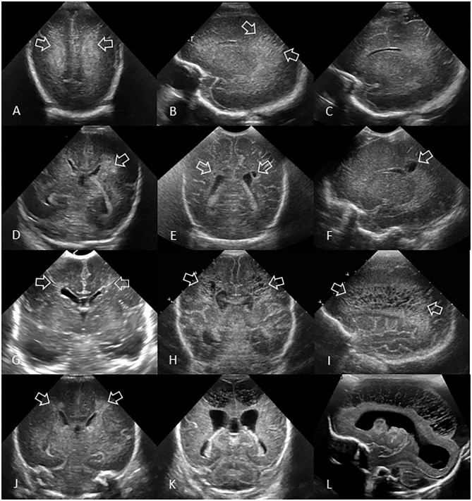
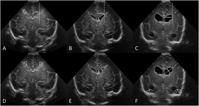
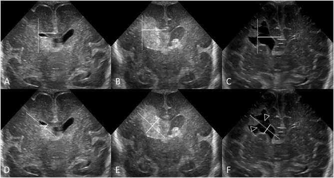
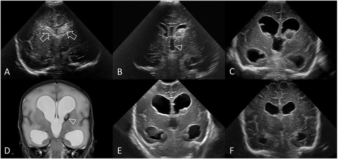
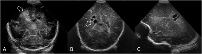
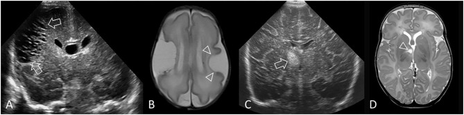
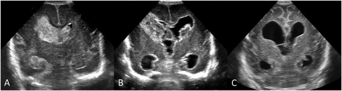
Acquired brain injury remains common in very preterm infants and is associated with significant risks for short- and long-term morbidities. Cranial ultrasound has been widely adopted as the first-line neuroimaging modality to study the neonatal brain. It can reliably detect clinically significant abnormalities that include germinal matrix and intraventricular hemorrhage, periventricular hemorrhagic infarction, post-hemorrhagic ventricular dilatation, cerebellar hemorrhage, and white matter injury. The purpose of this article is to provide a consensus approach for detecting and classifying preterm brain injury to reduce variability in diagnosis and classification between neonatologists and radiologists. Our overarching goal with this work was to achieve homogeneity between different neonatal intensive care units across a large country (Canada) with regards to classification, timing of brain injury screening and frequency of follow up imaging. We propose an algorithmic approach that can help stratify different grades of germinal matrix-intraventricular hemorrhage, white matter injury, and ventricular dilatation in very preterm infants.
获得性脑损伤在极早产儿中仍然很常见,并且与短期和长期发病的重大风险相关。头颅超声已被广泛用作研究新生儿脑的一线神经影像学检查方法。它能够可靠地检测出具有临床意义的异常情况,包括生发基质和脑室内出血、脑室周围出血性梗死、出血后脑室扩张、小脑出血和白质损伤。本文的目的是提供一种用于检测和分类早产儿脑损伤的共识方法,以减少新生儿科医生和放射科医生在诊断和分类上的差异。我们开展这项工作的总体目标是,在一个大国(加拿大)的不同新生儿重症监护病房之间,在脑损伤分类、筛查时间和后续影像学检查频率方面实现一致性。我们提出一种算法方法,该方法有助于对极早产儿生发基质 - 脑室内出血、白质损伤和脑室扩张的不同等级进行分层。