Zhang Yu, Hu Jun, Li Xiang, Qin Xiaodong
Department of Trauma, the First Affiliating Hospital of Nanjing Medical University & Jiangsu Province Hospital, 300 Guangzhou Road, Nanjing, 210029, China.
BMC Musculoskelet Disord. 2020 Mar 28;21(1):195. doi: 10.1186/s12891-020-03212-6.
To introduce an unreported intraoperative complication in intramedullary nailing (IN) of an anatomically reduced trochanteric fracture variant characterized by a basicervical fracture line and coronally disrupted greater trochanter (GT).
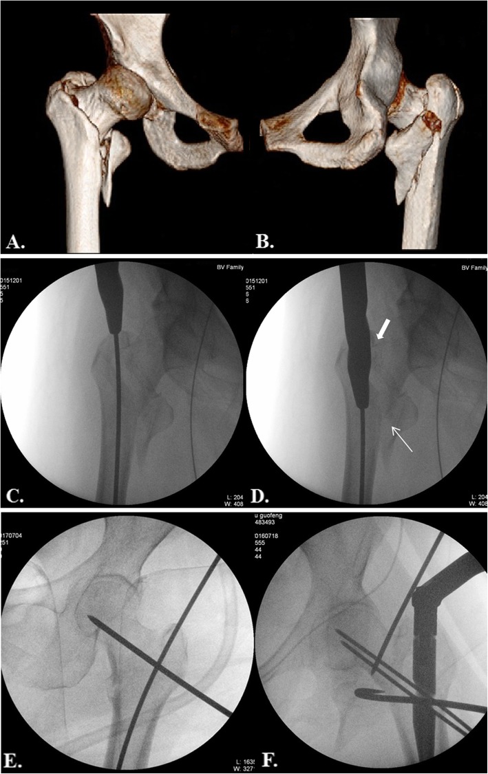
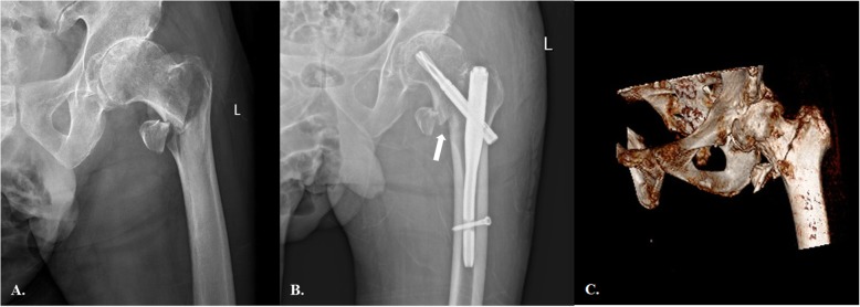
A total of 414 trochanteric fractures (TF) treated with intramedullary nails from 2013 to 2017 were included in this study. After analysis of intraoperative fluoroscopy data, 33 cases, including 21 females and 12 males, with a mean age of 72.5 years (33 to 96 years) were identified for internal rotation of the cephalocervical fragment and inferior opening at the basicervical fracture line caused by nailing a satisfactorily reduced TF. The morphological features of this group of patients were analyzed on computed tomography (CT) scan. On radiograph, the magnitude of the displacement and final femoral neck-shaft angle (NSA) were measured.
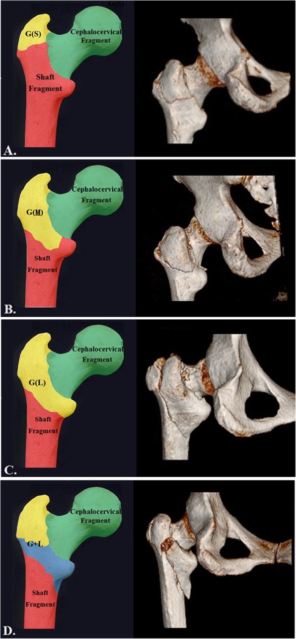
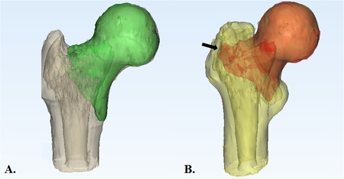
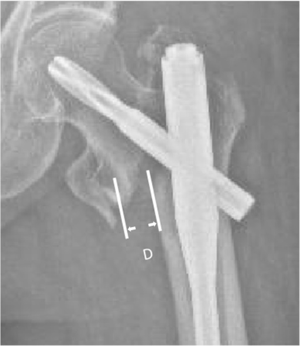
CT analysis demonstrated that the basicervical fracture line and the posterolateral fragment (PLF) detached from the GT were the two dominant features of this cohort. They were classified according to the number of main fragments: a 3-fragmentary subgroup containing three consistent fragments (cephalocervical fragment, PLF and distal femoral shaft) and a 4-fragmentary subgroup embracing one additional fragment (lesser trochanter). The four subtypes were as follows: the 3-fragmentary S indicating a small PLF (6 cases), the 3-fragmentary M presenting a moderate PLF (3 cases), the 3-fragmentary L standing for the PLF involving whole lesser trochanter (LT) (4 cases) and the 4-fragmentary GL incorporating separated PLF and LT fragments (20 cases). Geological analysis demonstrated that the majority of the basicervical fracture lines (81.8%) just crossed the center of the piriformis fossa, while the others marginally involved the medial wall of the GT. Postoperatively, the mean width of the inferior opening at the basicervical region was 9.2 ± 4.6 mm. The mean NSA was 135.2 ± 7.8 degrees. The comparison between the 3- and 4-fragmentary subgroups revealed no significant differences in magnitude of displacement and NSA.
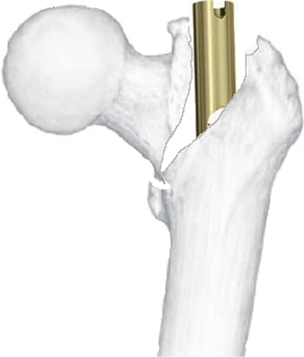
This unreported intraoperative complication predominantly occurred in the intramedullary nailed basicervical trochanteric fracture variant combined with a PLF from the GT. The magnitude of the secondary displacement was substantial and resulted in a relative valgus reduction. This secondary displacement was caused by an impingement of the reamer with the superolateral cortex of the cephalocervical fragment and should be addressed during the operation.
Therapy IV.
介绍一种未报道过的解剖复位型转子间骨折髓内钉固定术中并发症,其特征为基底部骨折线和冠状面移位的大转子(GT)。
本研究纳入了2013年至2017年采用髓内钉治疗的414例转子间骨折(TF)。分析术中透视数据后,确定33例患者(21例女性和12例男性),平均年龄72.5岁(33至96岁),这些患者在对满意复位的TF进行髓内钉固定后出现头颈骨块内旋和基底部骨折线下方开口。对这组患者的形态学特征进行了计算机断层扫描(CT)分析。在X线片上,测量移位程度和最终的股骨颈干角(NSA)。
CT分析表明,基底部骨折线和从GT分离的后外侧骨块(PLF)是该队列的两个主要特征。根据主要骨块数量进行分类:一个包含三个一致骨块(头颈骨块、PLF和股骨干远端)的三骨块亚组和一个包含一个额外骨块(小转子)的四骨块亚组。四种亚型如下:3 - 骨块S型表示小PLF(6例),3 - 骨块M型表示中等PLF(3例),3 - 骨块L型表示PLF累及整个小转子(LT)(4例),4 - 骨块GL型包含分离的PLF和LT骨块(20例)。地质分析表明,大多数基底部骨折线(81.8%)刚好穿过梨状窝中心,而其他骨折线则轻微累及GT的内侧壁。术后,基底部区域下方开口的平均宽度为9.2±4.6mm。平均NSA为135.2±7.8度。三骨块亚组和四骨块亚组之间在移位程度和NSA方面无显著差异。
这种未报道过的术中并发症主要发生在髓内钉固定的基底部转子间骨折变异型合并来自GT的PLF患者中。继发移位程度较大,导致相对外翻减小。这种继发移位是由扩髓器对头颈骨块上外侧皮质的撞击引起 的,手术中应予以处理。
治疗IV级。