Institute of Biology, Leiden University, 2311 EZ Leiden, The Netherlands.
Department of Molecular Biology, International Institute of Molecular and Cell Biology, 02-109 Warsaw, Poland.
Cells. 2020 Mar 26;9(4):797. doi: 10.3390/cells9040797.
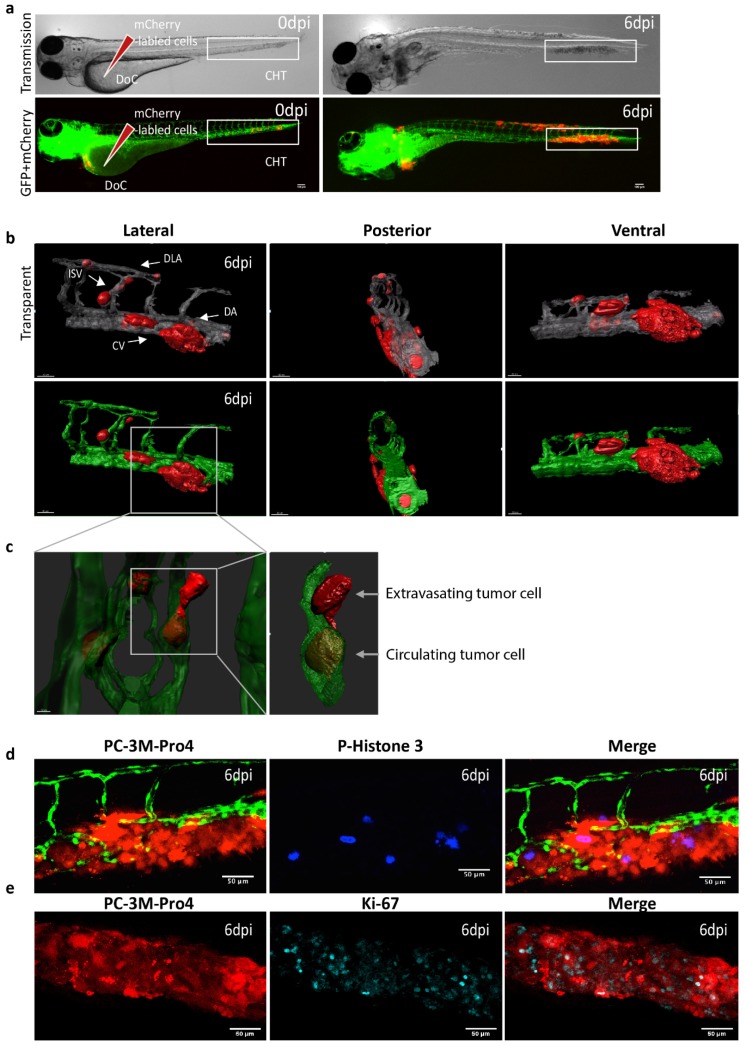
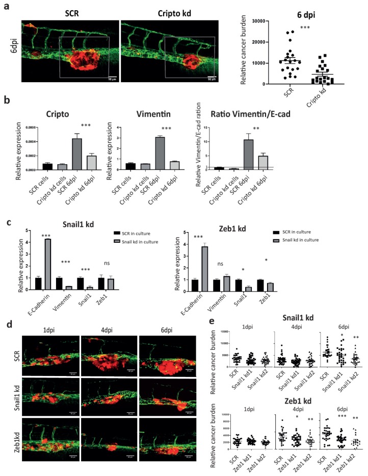
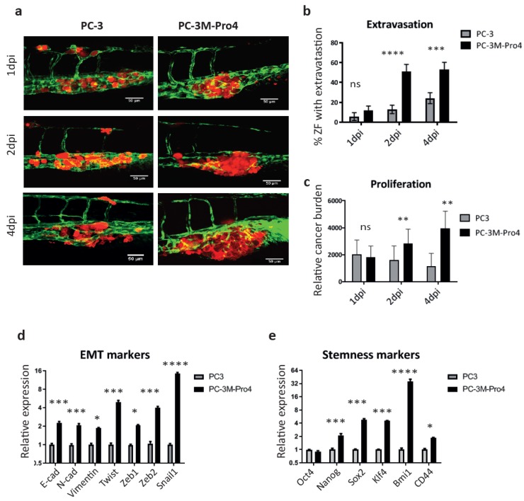
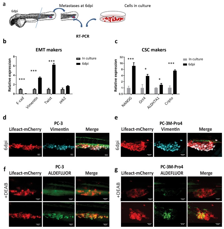
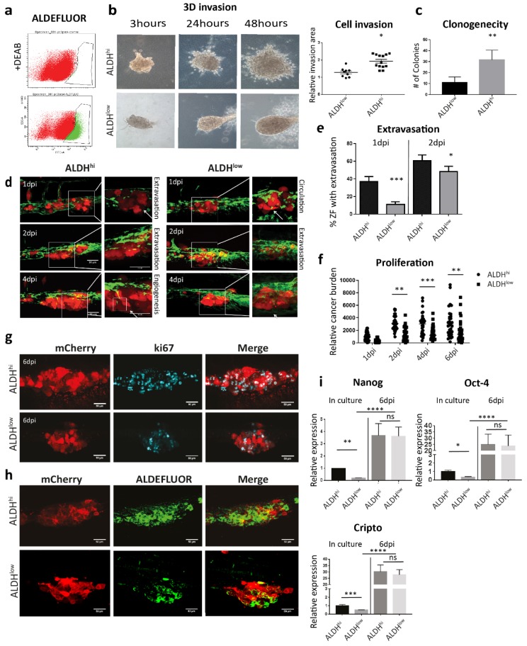
To visually and genetically trace single-cell dynamics of human prostate cancer (PCa) cells at the early stage of metastasis, a zebrafish (ZF) xenograft model was employed. The phenotypes of intravenously transplanted fluorescent cells were monitored by high-resolution, single-cell intravital confocal and light-sheet imaging. Engrafted osteotropic, androgen independent PCa cells were extravasated from caudle vein, invaded the neighboring tissue, proliferated and formed experimental metastases around caudal hematopoietic tissue (CHT) in four days. Gene expression comparison between cells in culture and in CHT revealed that engrafted PCa cells responded to the ZF microenvironment by elevating expression of epithelial-mesenchymal transition (EMT) and stemness markers. Next, metastatic potentials of ALDH cancer stem-like cells (CSCs) and ALDH non-CSCs were analyzed in ZF. Engraftment of CSCs induced faster metastatic onset, however after six days both cell subpopulations equally responded to the ZF microenvironment, resulting in the same increase of stemness genes expression including Nanog, Oct-4 and Cripto. Knockdown of Cripto significantly reduced the vimentin/E-cadherin ratio in engrafted cells, indicating that Cripto is required for transduction of the microenvironment signals from the ZF niche to increase mesenchymal potential of cells. Targeting of either Cripto or EMT transcriptional factors Snail 1 and Zeb1 significantly suppressed metastatic growth. These data indicated that zebrafish microenvironment governed the CSC/EMT plasticity of human PCa cells promoting metastasis initiation.
为了在人类前列腺癌(PCa)转移的早期阶段直观且基因上追踪单细胞的动态变化,我们采用了斑马鱼(ZF)异种移植模型。通过高分辨率、单细胞活体共聚焦和光片成像监测静脉内移植荧光细胞的表型。骨趋向性、雄激素非依赖性的 PCa 细胞从尾静脉逸出,侵袭邻近组织,在四天内在尾造血组织(CHT)周围增殖并形成实验性转移。细胞培养和 CHT 之间的基因表达比较表明,移植的 PCa 细胞通过上调上皮-间充质转化(EMT)和干细胞标记物来响应 ZF 微环境。接下来,在 ZF 中分析了具有醛脱氢酶(ALDH)的癌症干细胞样细胞(CSCs)和 ALDH 非-CSCs 的转移潜力。CSC 的移植诱导了更快的转移起始,然而在六天后,这两个细胞亚群对 ZF 微环境同样做出反应,导致包括 Nanog、Oct-4 和 Cripto 在内的干细胞基因表达相同增加。Cripto 的敲低显著降低了移植细胞中波形蛋白/E-钙黏蛋白的比值,表明 Cripto 是将 ZF 龛微环境信号转导到增加细胞间充质潜能所必需的。靶向 Cripto 或 EMT 转录因子 Snail 1 和 Zeb1 均可显著抑制转移生长。这些数据表明,斑马鱼微环境控制了人类 PCa 细胞的 CSC/EMT 可塑性,促进了转移的起始。