Department of Oral & Maxillofacial Surgery, University of Groningen, University Medical Center Groningen, the Netherlands.
Department of Gastroenterology and Hepatology, University of Groningen, University Medical Center Groningen, the Netherlands.
Theranostics. 2020 Mar 4;10(9):3994-4005. doi: 10.7150/thno.43227. eCollection 2020.
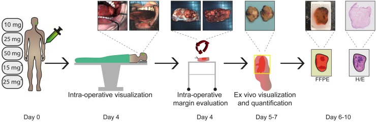
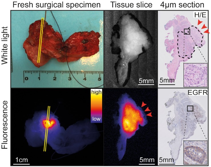
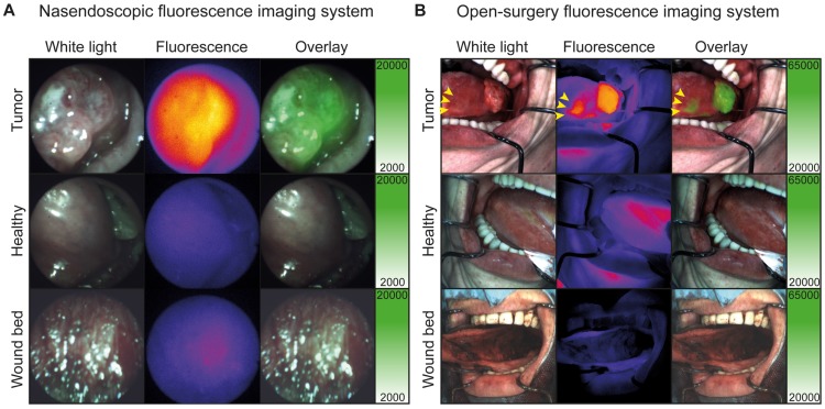
Tumor-positive resection margins are present in up to 23% of head and neck cancer (HNC) surgeries, as intraoperative techniques for evaluation of the resection margins are lacking. In this study, we investigated the safety and potential clinical value of fluorescence-guided imaging (FGI) for resection margin evaluation in HNC patients. We determined the optimal cetuximab-800CW dose by quantification of intrinsic fluorescence values using multi-diameter single-fiber reflectance, single-fiber fluorescence (MDSFR/SFF) spectroscopy. : Five cohorts of three HNC patients received cetuximab-800CW systemically: three single dose cohorts (10, 25, 50 mg) and two cohorts pre-dosed with 75 mg unlabeled cetuximab (15 or 25 mg). Fluorescence visualization and MDSFR/SFF spectroscopy quantification was performed and were correlated to histopathology. : There were no study-related adverse events higher than Common Terminology Criteria for Adverse Events grade-II. Quantification of intrinsic fluorescence values showed a dose-dependent increase in background fluorescence in the single dose cohorts (), which remained consistently low in the pre-dosed cohorts . Resection margin status was evaluated with a sensitivity of 100% (4/4 tumor-positive margins) and specificity of 91% (10/11 tumor-negative margins). : A pre-dose of 75 mg unlabeled cetuximab followed by 15 mg cetuximab-800CW was considered the optimal dose based on safety, fluorescence visualization and quantification of intrinsic fluorescence values. We were able to use a lower dose cetuximab-800CW than previously described, while remaining a high sensitivity for tumor detection due to application of equipment optimized for IRDye800CW detection, which was validated by quantification of intrinsic fluorescence values.
肿瘤阳性切缘见于多达 23%的头颈部癌症 (HNC) 手术中,因为术中评估切缘的技术缺乏。在这项研究中,我们研究了荧光引导成像 (FGI) 对头颈部癌症患者切缘评估的安全性和潜在临床价值。我们通过使用多直径单纤维反射率、单纤维荧光 (MDSFR/SFF) 光谱学来定量内源性荧光值来确定最佳西妥昔单抗-800CW 剂量。五组三名头颈部癌症患者接受了西妥昔单抗-800CW 系统治疗:三组单剂量组 (10、25、50mg) 和两组预剂量组,给予 75mg 未标记的西妥昔单抗 (15 或 25mg)。进行荧光可视化和 MDSFR/SFF 光谱学定量,并与组织病理学相关联。研究中没有比常见不良事件术语标准二级更高的不良事件。内源性荧光值的定量显示单剂量组的背景荧光呈剂量依赖性增加 (),而预剂量组的荧光一直保持较低水平。用 100%的敏感性 (4/4 个肿瘤阳性切缘) 和 91%的特异性 (10/11 个肿瘤阴性切缘) 评估切缘状态。基于安全性、荧光可视化和内源性荧光值的定量,我们认为 75mg 未标记的西妥昔单抗预剂量加 15mg 西妥昔单抗-800CW 是最佳剂量。由于应用了针对 IRDye800CW 检测进行了优化的设备,我们能够使用比以前描述的更低剂量的西妥昔单抗-800CW,同时由于内源性荧光值的定量验证,仍保持对肿瘤检测的高敏感性。