CAP‑Paris Tech., INSERM U1275, Lariboisière Hospital, 75010 Paris, France.
Oncol Rep. 2020 Jun;43(6):1797-1804. doi: 10.3892/or.2020.7572. Epub 2020 Apr 1.
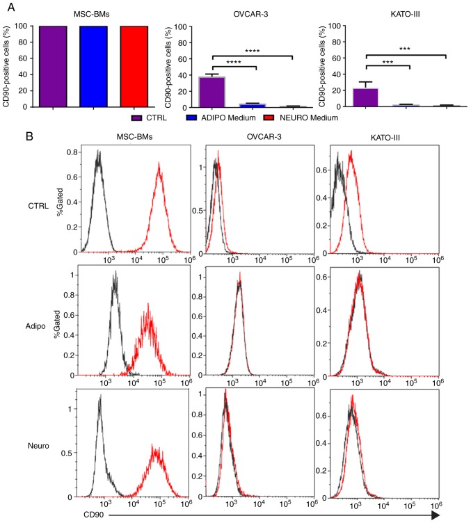
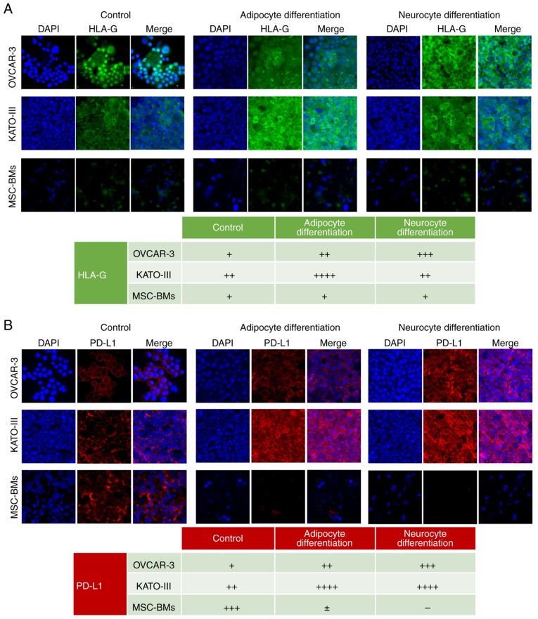
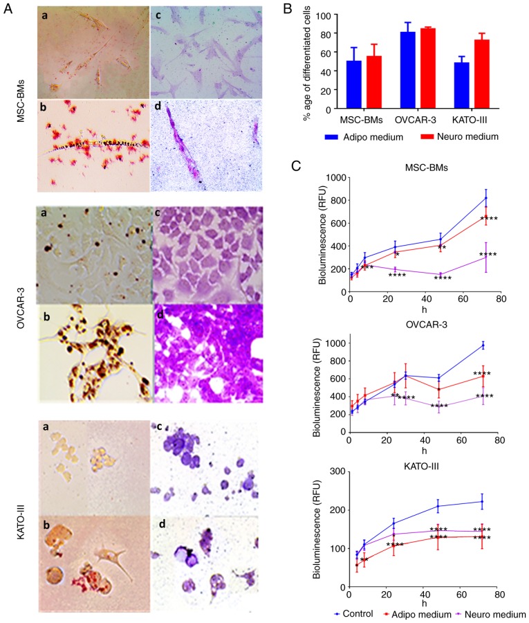
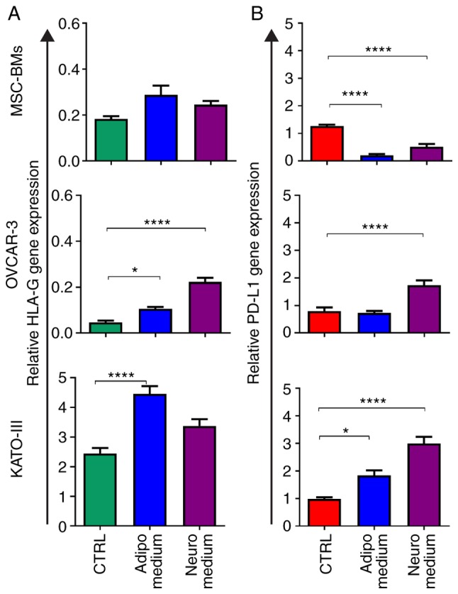
A tumor contains special types of cells that have characteristics similar to stem cells that aid in tumor initiation, evasion and proliferation and are often resistant to chemotherapy. These cancer stem cells can be differentiated to eradicate their stemness and proliferative capacity by differentiating agents. This study investigated the effect of differentiation on the expression of two immune checkpoint inhibitors, human leukocyte antigen‑G (HLA‑G) and programmed death ligand‑1 (PD‑L1). Two cancer cell lines (OVCAR‑3‑NIH and KATO‑III) were treated with adipocyte and neurocyte differentiation media for 14 days. Bone‑marrow derived mesenchymal stem cells (BM‑MSCs) were used as control healthy stem cells. We found that the cancer cell lines (OVCAR‑3‑NIH and KATO‑III) when subjected to differentiation lost their proliferation ability. BM‑MSC proliferation was not halted but was decreased in the adipocyte differentiation media. There was no decrease in the CD90 stem cell marker in the BM‑MSCs; however, both cancer cell lines showed decreased CD90 stem cell marker. A significant increase in HLA‑G was noted for both the cancer cell lines following adipocyte differentiation. No effect was found for BM‑MSCs. Moreover, an increase in PD‑L1 in cancer cell lines was found following neurocyte differentiation. Moreover, we found that differentiation resulted in decreased PD‑L1 expression in BM‑MSCs. Differentiation therapy of cancer stem cells may result in increased immunosuppression ability, hence causing hindrance in the removal of cancer cells. Moreover, the differentiation of healthy stem cells can result in increased immunogenic reactivity owing to a decrease in PD‑L1 expression.
肿瘤包含具有与有助于肿瘤起始、逃逸和增殖的干细胞相似特征的特殊类型细胞,并且往往对化疗具有抗性。这些癌症干细胞可以通过分化剂分化来消除其干性和增殖能力。本研究调查了分化对两种免疫检查点抑制剂人白细胞抗原 G (HLA-G)和程序性死亡配体 1 (PD-L1)表达的影响。将两种癌细胞系 (OVCAR-3-NIH 和 KATO-III) 用脂肪细胞和神经细胞分化培养基处理 14 天。骨髓间充质干细胞 (BM-MSCs) 用作对照健康干细胞。我们发现,癌细胞系 (OVCAR-3-NIH 和 KATO-III) 在分化时失去了增殖能力。BM-MSC 的增殖没有停止,但在脂肪细胞分化培养基中减少。BM-MSCs 中的 CD90 干细胞标志物没有减少;然而,两种癌细胞系均显示 CD90 干细胞标志物减少。脂肪细胞分化后,两种癌细胞系的 HLA-G 均显著增加。BM-MSCs 未发现此作用。此外,神经细胞分化后,癌细胞系中 PD-L1 增加。此外,我们发现分化导致 BM-MSCs 中 PD-L1 表达减少。癌症干细胞的分化治疗可能导致免疫抑制能力增加,从而阻碍癌细胞的清除。此外,由于 PD-L1 表达减少,健康干细胞的分化可能导致免疫原性反应性增加。