• 文献检索
  • 文档翻译
  • 深度研究
  • 学术资讯
  • Suppr Zotero 插件Zotero 插件
  • 邀请有礼
  • 套餐&价格
  • 历史记录
应用&插件
Suppr Zotero 插件Zotero 插件浏览器插件Mac 客户端Windows 客户端微信小程序
定价
高级版会员购买积分包购买API积分包
服务
文献检索文档翻译深度研究API 文档MCP 服务
关于我们
关于 Suppr公司介绍联系我们用户协议隐私条款
关注我们

Suppr 超能文献

核心技术专利:CN118964589B侵权必究
粤ICP备2023148730 号-1Suppr @ 2026

文献检索

告别复杂PubMed语法,用中文像聊天一样搜索,搜遍4000万医学文献。AI智能推荐,让科研检索更轻松。

立即免费搜索

文件翻译

保留排版,准确专业,支持PDF/Word/PPT等文件格式,支持 12+语言互译。

免费翻译文档

深度研究

AI帮你快速写综述,25分钟生成高质量综述,智能提取关键信息,辅助科研写作。

立即免费体验

用分形分析评估短种植体周围骨小梁微观结构变化

Evaluation of the peri-implant bone trabecular microstructure changes in short implants with fractal analysis.

作者信息

Kış Hatice Cansu, Güleryüz Gürbulak Ayşegül

机构信息

Department of Oral and Maxillofacial Radiology, Faculty of Dentistry, Nuh Naci Yazgan University, Kayseri, Turkey.

Department of Prosthetic Dentistry, Faculty of Dentistry, Erciyes University, Kayseri, Turkey.

出版信息

Int J Implant Dent. 2020 Apr 1;6(1):13. doi: 10.1186/s40729-020-00209-7.

DOI:10.1186/s40729-020-00209-7
PMID:32236773
原文链接:https://pmc.ncbi.nlm.nih.gov/articles/PMC7109216/
Abstract

BACKGROUNDS

This study aimed to evaluate the microstructural changes in the peri-implant bone in patients with short implants in terms of implant survival status by using fractal analysis measurements.

RESULTS

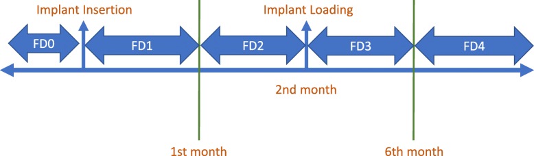
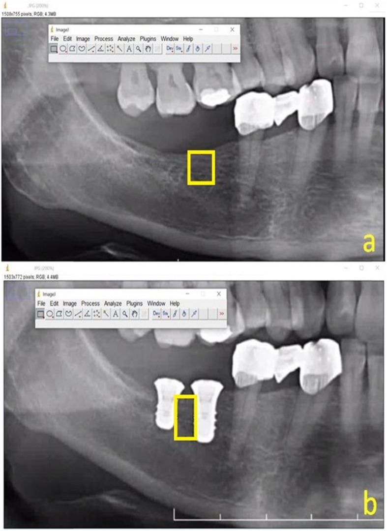
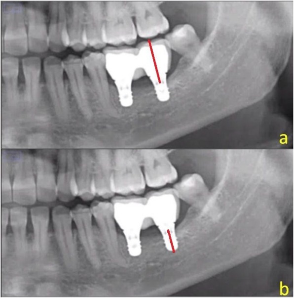
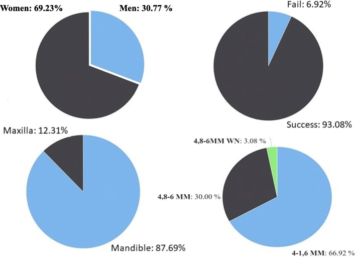
Dental panoramic radiographs (DPRs) of 67 patients were examined and included in this study. Fractal analysis and measurement of the crown-implant ratio were performed with ImageJ. The fractal analysis measurement was performed on the DPRs obtained at preoperative (FD0) and in the follow-up periods (after 2 ± 2 weeks (FD1), 2 months ± 2 weeks (FD2), 6 months ± 2 weeks (FD3), and 12 months + (FD4)). A p value < 0.05 was considered statistically significant. Power analyses were conducted for the test results that did not reject null hypothesis. A significant difference was found in the FD1 and FD2 values between the implant survival groups (p < 0.001 and p = 0.023, respectively). The mean FD1 and FD2 values of the success group were significantly higher than those of the failure group.

CONCLUSIONS

Fractal analysis is a useful method to measure the trabecular microstructure of bone in non-standardized dental radiographs. The present study has a low power to reject the null hypothesis because of the low number of cases of implant failure. Therefore, further study with a large sample size is warranted. In clinical practice, the survival of implants may be predicted by analyzing fractal dimension of the surrounding trabecular bone of the implants.

摘要

背景

本研究旨在通过分形分析测量,根据种植体存活状态评估短种植体患者种植体周围骨的微观结构变化。

结果

对67例患者的牙科全景X线片(DPR)进行了检查并纳入本研究。使用ImageJ进行分形分析和冠种植体比率测量。在术前(FD0)以及随访期(2±2周后(FD1)、2个月±2周(FD2)、6个月±2周(FD3)和12个月及以后(FD4))获得的DPR上进行分形分析测量。p值<0.05被认为具有统计学意义。对未拒绝原假设的测试结果进行功效分析。在种植体存活组之间,FD1和FD2值存在显著差异(分别为p<0.001和p = 0.023)。成功组的平均FD1和FD2值显著高于失败组。

结论

分形分析是测量非标准化牙科X线片中骨小梁微观结构的有用方法。由于种植体失败病例数较少,本研究拒绝原假设的功效较低。因此,有必要进行更大样本量的进一步研究。在临床实践中,可通过分析种植体周围骨小梁的分形维数来预测种植体的存活情况。

https://cdn.ncbi.nlm.nih.gov/pmc/blobs/4152/7109216/690d66e9d4c7/40729_2020_209_Fig7_HTML.jpg
https://cdn.ncbi.nlm.nih.gov/pmc/blobs/4152/7109216/6c8e38230424/40729_2020_209_Fig1_HTML.jpg
https://cdn.ncbi.nlm.nih.gov/pmc/blobs/4152/7109216/49866f294bb4/40729_2020_209_Fig2_HTML.jpg
https://cdn.ncbi.nlm.nih.gov/pmc/blobs/4152/7109216/7a17262dbb56/40729_2020_209_Fig3_HTML.jpg
https://cdn.ncbi.nlm.nih.gov/pmc/blobs/4152/7109216/20a1db47b3b8/40729_2020_209_Fig4_HTML.jpg
https://cdn.ncbi.nlm.nih.gov/pmc/blobs/4152/7109216/5f795b479314/40729_2020_209_Fig5_HTML.jpg
https://cdn.ncbi.nlm.nih.gov/pmc/blobs/4152/7109216/a216cc983130/40729_2020_209_Fig6_HTML.jpg
https://cdn.ncbi.nlm.nih.gov/pmc/blobs/4152/7109216/690d66e9d4c7/40729_2020_209_Fig7_HTML.jpg

相似文献

1
Evaluation of the peri-implant bone trabecular microstructure changes in short implants with fractal analysis.用分形分析评估短种植体周围骨小梁微观结构变化
Int J Implant Dent. 2020 Apr 1;6(1):13. doi: 10.1186/s40729-020-00209-7.
2
Fractal analysis for the assessment of trabecular peri-implant alveolar bone using panoramic radiographs.使用全景X线片进行分形分析以评估种植体周围牙槽骨小梁
Clin Oral Investig. 2015 Mar;19(2):519-24. doi: 10.1007/s00784-014-1245-y. Epub 2014 May 7.
3
Effect of a laser-ablated micron-scale modification of dental implant collar surface on changes in the vertical and fractal dimensions of peri-implant trabecular bone.激光消融对牙种植体颈部表面进行微米级改性对种植体周围小梁骨垂直维度和分形维数变化的影响。
Clin Ter. 2020 Sep-Oct;171(5):e385-e392. doi: 10.7417/CT.2020.2245.
4
In vivo assessment of trabecular bone structure using fractal analysis of distal radius radiographs.使用桡骨远端X线片的分形分析对小梁骨结构进行体内评估。
Med Phys. 2000 Nov;27(11):2594-9. doi: 10.1118/1.1319375.
5
Correlation of Fractal Dimension Values with Implant Insertion Torque and Resonance Frequency Values at Implant Recipient Sites.种植体植入部位的分形维数值与种植体植入扭矩及共振频率值的相关性。
Int J Oral Maxillofac Implants. 2016 Jan-Feb;31(1):55-62. doi: 10.11607/jomi.3965. Epub 2015 Oct 16.
6
Changes in the fractal dimension of peri-implant trabecular bone after loading: a retrospective study.加载后种植体周围松质骨分形维数的变化:一项回顾性研究。
J Periodontal Implant Sci. 2013 Oct;43(5):209-14. doi: 10.5051/jpis.2013.43.5.209. Epub 2013 Oct 29.
7
A clinical study of alveolar bone quality using the fractal dimension and the implant stability quotient.一项使用分形维数和种植体稳定性商数评估牙槽骨质量的临床研究。
J Periodontal Implant Sci. 2010 Feb;40(1):19-24. doi: 10.5051/jpis.2010.40.1.19. Epub 2010 Feb 28.
8
Evaluation of change in trabecular bone structure surrounding dental implants by fractal dimension analysis and comparison with radiomorphometric indicators: a retrospective study.基于分形维数分析评估种植体周围小梁骨结构的变化,并与放射形态计量学指标进行比较:一项回顾性研究。
PeerJ. 2022 Mar 22;10:e13145. doi: 10.7717/peerj.13145. eCollection 2022.
9
Damping factor for monitoring the bone interface at dental implants.用于监测牙种植体骨界面的阻尼因子。
Clin Oral Implants Res. 2007 Dec;18(6):738-42. doi: 10.1111/j.1600-0501.2007.01412.x. Epub 2007 Sep 20.
10
The use of fractal analysis to reveal remodelling in human alveolar bone following the placement of dental implants.利用分形分析揭示牙种植体植入后人类牙槽骨的重塑情况。
Arch Oral Biol. 1995 Jan;40(1):61-72. doi: 10.1016/0003-9969(94)00138-2.

引用本文的文献

1
Decoding dental images: a comprehensive review of fractal analysis.解读牙科影像:分形分析的全面综述
BDJ Open. 2025 Aug 21;11(1):74. doi: 10.1038/s41405-025-00365-1.
2
Automated fractal analysis for mandibular bone evaluation in type 1 diabetes mellitus using a novel single click approach.使用新型一键式方法对1型糖尿病患者下颌骨进行自动分形分析以进行评估。
Sci Rep. 2025 Aug 18;15(1):30266. doi: 10.1038/s41598-025-07464-x.
3
Evaluation of the bone structure surrounding photofunctionalized implants using the fractal analysis method: a split-mouth randomized clinical study.

本文引用的文献

1
The evaluation of the mandibular bone structure changes related to lactation with fractal analysis.应用分形分析评价与哺乳相关的下颌骨结构变化。
Oral Radiol. 2020 Jul;36(3):238-247. doi: 10.1007/s11282-019-00400-6. Epub 2019 Jul 27.
2
Fractal analysis for the assessment of trabecular peri-implant alveolar bone using panoramic radiographs.使用全景X线片进行分形分析以评估种植体周围牙槽骨小梁
Clin Oral Investig. 2015 Mar;19(2):519-24. doi: 10.1007/s00784-014-1245-y. Epub 2014 May 7.
3
Changes in the fractal dimension of peri-implant trabecular bone after loading: a retrospective study.
使用分形分析方法评估光功能化种植体周围的骨结构:一项双侧随机临床研究。
BMC Oral Health. 2025 Jul 1;25(1):964. doi: 10.1186/s12903-025-06330-6.
4
Evaluation of the long-term structural change caused by root canal treatment in the bone around the tooth by fractal analysis.通过分形分析评估根管治疗对牙齿周围骨组织造成的长期结构变化。
Oral Radiol. 2025 May 6. doi: 10.1007/s11282-025-00825-2.
5
Post-Extraction Bone Changes in Molars Within Personalized Implant-Prosthetic Therapy as Evaluated with Fractal Analysis of CBCT.通过锥形束计算机断层扫描(CBCT)的分形分析评估个性化种植修复治疗中磨牙拔牙后的骨变化。
J Pers Med. 2025 Apr 16;15(4):154. doi: 10.3390/jpm15040154.
6
Palatal rugae change shape following orthodontic treatment: a comparison between extraction and non-extraction borderline cases using fractal analysis and 3D superimposition.正畸治疗后腭皱襞形状发生改变:采用分形分析和三维叠加法对拔牙和非拔牙临界病例的比较
Eur J Orthod. 2024 Dec 4;47(1). doi: 10.1093/ejo/cjae070.
7
Fractal dimension, lacunarity, and bone area fraction analysis of peri-implant trabecular bone after prosthodontic loading.义齿修复加载后种植体周围松质骨的分形维数、孔隙度和骨面积分数分析
Oral Radiol. 2025 Jan;41(1):120-130. doi: 10.1007/s11282-024-00784-0. Epub 2024 Nov 11.
8
Pilot study of fractal dimension analysis of osteogenesis for bone substitute materials of Bio-Oss in lateral sinus augmentation.外侧窦提升中 Bio-Oss 骨替代材料成骨的分形维数分析的初步研究。
PLoS One. 2023 Dec 29;18(12):e0296248. doi: 10.1371/journal.pone.0296248. eCollection 2023.
9
Novel method to delineate palatal rugae and assess their complexity using fractal analysis.使用分形分析对腭皱襞进行描绘和评估其复杂性的新方法。
Sci Rep. 2022 Dec 16;12(1):21749. doi: 10.1038/s41598-022-25910-y.
10
Differentiation of osteosarcoma from osteomyelitis using microarchitectural analysis on panoramic radiographs.利用曲面体层片的微结构分析对骨肉瘤与骨髓炎进行鉴别诊断。
Sci Rep. 2022 Jul 19;12(1):12339. doi: 10.1038/s41598-022-16504-9.
加载后种植体周围松质骨分形维数的变化:一项回顾性研究。
J Periodontal Implant Sci. 2013 Oct;43(5):209-14. doi: 10.5051/jpis.2013.43.5.209. Epub 2013 Oct 29.
4
Diagnostic imaging of trabecular bone microstructure for oral implants: a literature review.口腔种植体小梁骨结构的影像学诊断:文献综述。
Dentomaxillofac Radiol. 2013;42(3):20120075. doi: 10.1259/dmfr.20120075.
5
Image resolution and exposure time of digital radiographs affects fractal dimension of periapical bone.数字化根尖片的图像分辨率和曝光时间会影响根尖周骨的分形维数。
Clin Oral Investig. 2012 Oct;16(5):1507-10. doi: 10.1007/s00784-011-0639-3. Epub 2011 Nov 29.
6
Fractal analysis of mandibular trabecular bone: optimal tile sizes for the tile counting method.下颌骨小梁骨的分形分析:瓦片计数法的最佳瓦片尺寸
Imaging Sci Dent. 2011 Jun;41(2):71-8. doi: 10.5624/isd.2011.41.2.71. Epub 2011 Jun 23.
7
Fractals in dentistry.牙科中的分形
J Dent. 2011 Apr;39(4):273-92. doi: 10.1016/j.jdent.2011.01.010. Epub 2011 Feb 15.
8
Peri-implant bone tissue assessment by comparing the outcome of intra-oral radiograph and cone beam computed tomography analyses to the histological standard.通过比较口腔内射线照相和锥形束计算机断层扫描分析的结果与组织学标准来评估种植体周围骨组织。
Clin Oral Implants Res. 2011 May;22(5):492-9. doi: 10.1111/j.1600-0501.2010.02029.x. Epub 2010 Dec 9.
9
Fractal analysis of dental radiographs to detect periodontitis-induced trabecular changes.用于检测牙周炎引起的骨小梁变化的牙科X光片分形分析
J Periodontal Res. 2008 Dec;43(6):658-64. doi: 10.1111/j.1600-0765.2007.01056.x. Epub 2008 Jun 24.
10
Technical factors in fractal analysis of periapical radiographs.根尖片分形分析中的技术因素。
Dentomaxillofac Radiol. 2006 Nov;35(6):393-7. doi: 10.1259/dmfr/30969642.