• 文献检索
  • 文档翻译
  • 深度研究
  • 学术资讯
  • Suppr Zotero 插件Zotero 插件
  • 邀请有礼
  • 套餐&价格
  • 历史记录
应用&插件
Suppr Zotero 插件Zotero 插件浏览器插件Mac 客户端Windows 客户端微信小程序
定价
高级版会员购买积分包购买API积分包
服务
文献检索文档翻译深度研究API 文档MCP 服务
关于我们
关于 Suppr公司介绍联系我们用户协议隐私条款
关注我们

Suppr 超能文献

核心技术专利:CN118964589B侵权必究
粤ICP备2023148730 号-1Suppr @ 2026

文献检索

告别复杂PubMed语法,用中文像聊天一样搜索,搜遍4000万医学文献。AI智能推荐,让科研检索更轻松。

立即免费搜索

文件翻译

保留排版,准确专业,支持PDF/Word/PPT等文件格式,支持 12+语言互译。

免费翻译文档

深度研究

AI帮你快速写综述,25分钟生成高质量综述,智能提取关键信息,辅助科研写作。

立即免费体验

LCK 表达可作为一种潜在的生物标志物,用于区分原发性中枢神经系统淋巴瘤和多形性胶质母细胞瘤。

LCK expression is a potential biomarker for distinguishing primary central nervous system lymphoma from glioblastoma multiforme.

机构信息

Tianjin Neurosurgical Institute, Tianjin Key Laboratory of Cerebrovascular and Neurodegenerative Diseases, Tianjin Huanhu Hospital, China.

出版信息

FEBS Open Bio. 2020 May;10(5):904-911. doi: 10.1002/2211-5463.12849. Epub 2020 Apr 13.

DOI:10.1002/2211-5463.12849
PMID:32237064
原文链接:https://pmc.ncbi.nlm.nih.gov/articles/PMC7193164/
Abstract

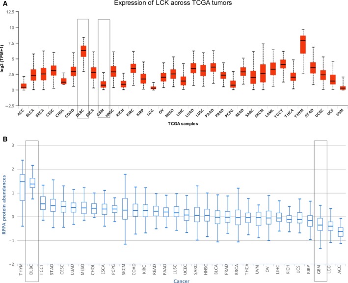
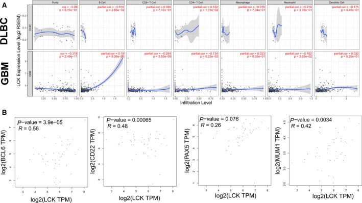
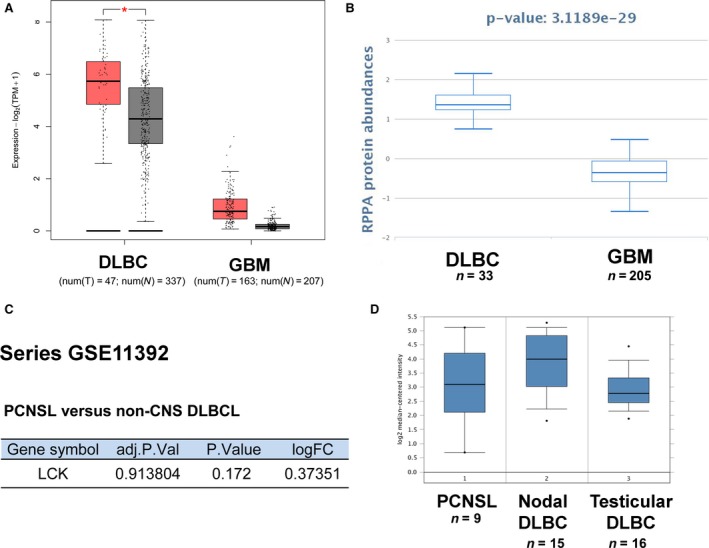
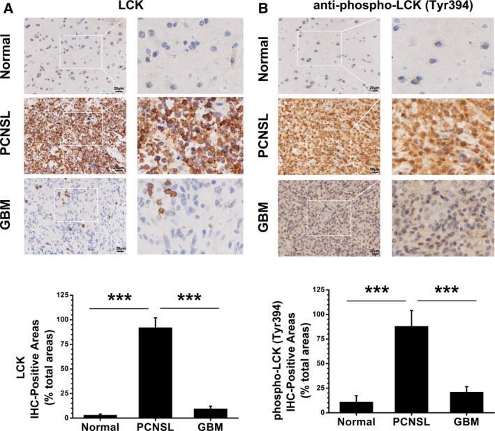
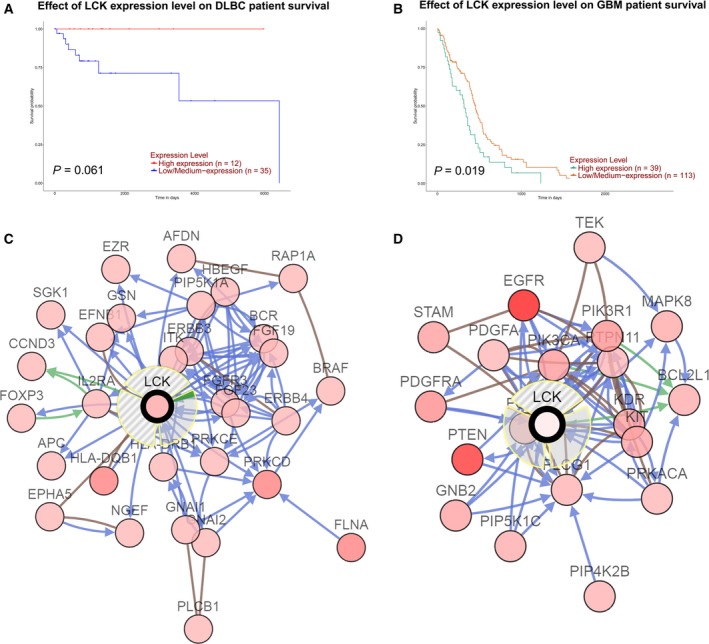
Glioblastoma multiforme (GBM) and primary central nervous system lymphoma (PCNSL) are both malignant cerebral tumors; however, their treatments are vastly different. Early and precise diagnosis is vital for subsequent adequate treatment to improve prognosis. Reliable biomarkers that can easily distinguish GBM and PCNSL are urgently needed. We evaluated the diagnostic potential of lymphocyte-specific protein tyrosine kinase (LCK) as a biomarker in differentiating PCNSL from GBM using established computational approaches (Gene Expression Profiling Interactive Analysis, The Cancer Proteome Atlas, Tumor Immune Estimation Resource, GEO, Oncomine) and immunohistochemistry. The results showed that LCK was expressed at a high level in PCNSL patients but at a low level in GBM patients. Moreover, LCK expression positively correlated with the levels of infiltrating B cells in diffuse large B-cell lymphoma (DLBCL) and GBM. Overall, bioinformatics analysis and immunohistochemistry revealed that LCK expression is a potential biomarker for distinguishing PCNSL from GBM.

摘要

多形性胶质母细胞瘤(GBM)和原发性中枢神经系统淋巴瘤(PCNSL)均为恶性脑肿瘤,但它们的治疗方法却大不相同。早期和准确的诊断对于后续的充分治疗以改善预后至关重要。目前急需可靠的生物标志物来区分 GBM 和 PCNSL。我们使用已建立的计算方法(基因表达谱交互式分析、癌症蛋白质组图谱、肿瘤免疫估计资源、GEO、Oncomine)和免疫组织化学评估了淋巴细胞特异性蛋白酪氨酸激酶(LCK)作为区分 PCNSL 和 GBM 的生物标志物的诊断潜力。结果表明,LCK 在 PCNSL 患者中表达水平较高,而在 GBM 患者中表达水平较低。此外,LCK 表达与弥漫性大 B 细胞淋巴瘤(DLBCL)和 GBM 中浸润 B 细胞的水平呈正相关。总的来说,生物信息学分析和免疫组织化学表明,LCK 表达是区分 PCNSL 和 GBM 的潜在生物标志物。

https://cdn.ncbi.nlm.nih.gov/pmc/blobs/3b59/7193164/6a19b0da2752/FEB4-10-904-g005.jpg
https://cdn.ncbi.nlm.nih.gov/pmc/blobs/3b59/7193164/aad5a7747fb4/FEB4-10-904-g001.jpg
https://cdn.ncbi.nlm.nih.gov/pmc/blobs/3b59/7193164/a11cf9d56f95/FEB4-10-904-g002.jpg
https://cdn.ncbi.nlm.nih.gov/pmc/blobs/3b59/7193164/c0c58b4b9f42/FEB4-10-904-g003.jpg
https://cdn.ncbi.nlm.nih.gov/pmc/blobs/3b59/7193164/58399e3d0648/FEB4-10-904-g004.jpg
https://cdn.ncbi.nlm.nih.gov/pmc/blobs/3b59/7193164/6a19b0da2752/FEB4-10-904-g005.jpg

相似文献

1
LCK expression is a potential biomarker for distinguishing primary central nervous system lymphoma from glioblastoma multiforme.LCK 表达可作为一种潜在的生物标志物,用于区分原发性中枢神经系统淋巴瘤和多形性胶质母细胞瘤。
FEBS Open Bio. 2020 May;10(5):904-911. doi: 10.1002/2211-5463.12849. Epub 2020 Apr 13.
2
Diagnostic delay and prognosis in primary central nervous system lymphoma compared with glioblastoma multiforme.原发性中枢神经系统淋巴瘤与多形性胶质母细胞瘤的诊断延迟及预后比较。
Neurol Sci. 2016 Jan;37(1):23-29. doi: 10.1007/s10072-015-2353-4. Epub 2015 Aug 2.
3
Clinicopathological analysis and specific discriminating markers of interleukin detection in cerebrospinal fluid with primary central nervous system lymphoma: results from a retrospective study.原发性中枢神经系统淋巴瘤患者脑脊液中白细胞介素检测的临床病理分析及特异性鉴别标志物:一项回顾性研究结果。
Ann Hematol. 2023 Aug;102(8):2153-2163. doi: 10.1007/s00277-023-05301-7. Epub 2023 Jun 8.
4
F-FDG PET/CT in immunocompetent patients with primary central nervous system lymphoma: Differentiation from glioblastoma and correlation with DWI.免疫功能正常的原发性中枢神经系统淋巴瘤患者的 F-FDG PET/CT:与胶质母细胞瘤的鉴别诊断及与 DWI 的相关性。
Eur J Radiol. 2018 Jul;104:26-32. doi: 10.1016/j.ejrad.2018.04.020. Epub 2018 Apr 22.
5
MR imaging based fractal analysis for differentiating primary CNS lymphoma and glioblastoma.基于磁共振成像的分形分析在原发性中枢神经系统淋巴瘤和胶质母细胞瘤鉴别诊断中的应用。
Eur Radiol. 2019 Mar;29(3):1348-1354. doi: 10.1007/s00330-018-5658-x. Epub 2018 Aug 30.
6
Intraoperative Flow Cytometry Enables the Differentiation of Primary Central Nervous System Lymphoma from Glioblastoma.术中流式细胞术有助于鉴别原发性中枢神经系统淋巴瘤和胶质母细胞瘤。
World Neurosurg. 2018 Apr;112:e261-e268. doi: 10.1016/j.wneu.2018.01.033. Epub 2018 Jan 9.
7
Glioblastoma and primary central nervous system lymphoma: Preoperative differentiation by using MRI-based 3D texture analysis.胶质母细胞瘤和原发性中枢神经系统淋巴瘤:基于MRI的三维纹理分析术前鉴别诊断
Clin Neurol Neurosurg. 2018 Oct;173:84-90. doi: 10.1016/j.clineuro.2018.08.004. Epub 2018 Aug 2.
8
Array-based profiling of the lymphoma cell DNA methylome does not unequivocally distinguish primary lymphomas of the central nervous system from non-CNS diffuse large B-cell lymphomas.基于阵列的淋巴瘤细胞 DNA 甲基化组分析不能明确区分中枢神经系统原发性淋巴瘤和非中枢神经系统弥漫性大 B 细胞淋巴瘤。
Genes Chromosomes Cancer. 2019 Jan;58(1):66-69. doi: 10.1002/gcc.22687. Epub 2018 Nov 29.
9
Primary central nervous system lymphoma and atypical glioblastoma: differentiation using the initial area under the curve derived from dynamic contrast-enhanced MR and the apparent diffusion coefficient.原发中枢神经系统淋巴瘤和非典型性脑胶质瘤:基于动态对比增强磁共振的初始曲线下面积和表观扩散系数的鉴别诊断。
Eur Radiol. 2017 Apr;27(4):1344-1351. doi: 10.1007/s00330-016-4484-2. Epub 2016 Jul 19.
10
Presence of apoptosis distinguishes primary central nervous system lymphoma from glioblastoma during intraoperative consultation.在术中会诊时,细胞凋亡的存在可将原发性中枢神经系统淋巴瘤与胶质母细胞瘤区分开来。
Clin Neuropathol. 2018 May/Jun;37(3):105-111. doi: 10.5414/NP301075.

引用本文的文献

1
Development and validation of a glioma prognostic model based on telomere-related genes and immune infiltration analysis.基于端粒相关基因和免疫浸润分析的胶质瘤预后模型的构建与验证
Transl Cancer Res. 2024 Jul 31;13(7):3182-3199. doi: 10.21037/tcr-23-2294. Epub 2024 Jul 22.
2
Small Molecule Tyrosine Kinase Inhibitors (TKIs) for Glioblastoma Treatment.小分子酪氨酸激酶抑制剂(TKIs)治疗脑胶质瘤。
Int J Mol Sci. 2024 Jan 23;25(3):1398. doi: 10.3390/ijms25031398.
3
New horizons in drug discovery of lymphocyte-specific protein tyrosine kinase (Lck) inhibitors: a decade review (2011-2021) focussing on structure-activity relationship (SAR) and docking insights.

本文引用的文献

1
Beyond TCR Signaling: Emerging Functions of Lck in Cancer and Immunotherapy.超越 TCR 信号:Lck 在癌症和免疫疗法中的新兴功能。
Int J Mol Sci. 2019 Jul 16;20(14):3500. doi: 10.3390/ijms20143500.
2
Regulation of human glioma cell migration, tumor growth, and stemness gene expression using a Lck targeted inhibitor.利用 Lck 靶向抑制剂调控人神经胶质瘤细胞迁移、肿瘤生长和干性基因表达。
Oncogene. 2019 Mar;38(10):1734-1750. doi: 10.1038/s41388-018-0546-z. Epub 2018 Oct 23.
3
The Ciliary Machinery Is Repurposed for T Cell Immune Synapse Trafficking of LCK.
淋巴细胞特异性蛋白酪氨酸激酶 (Lck) 抑制剂药物发现的新领域:十年综述 (2011-2021),重点关注结构-活性关系 (SAR) 和对接见解。
J Enzyme Inhib Med Chem. 2021 Dec;36(1):1574-1602. doi: 10.1080/14756366.2021.1937143.
纤毛机械被重新用于 LCK 的 T 细胞免疫突触运输。
Dev Cell. 2018 Oct 8;47(1):122-132.e4. doi: 10.1016/j.devcel.2018.08.012. Epub 2018 Sep 13.
4
MR imaging based fractal analysis for differentiating primary CNS lymphoma and glioblastoma.基于磁共振成像的分形分析在原发性中枢神经系统淋巴瘤和胶质母细胞瘤鉴别诊断中的应用。
Eur Radiol. 2019 Mar;29(3):1348-1354. doi: 10.1007/s00330-018-5658-x. Epub 2018 Aug 30.
5
TIMER: A Web Server for Comprehensive Analysis of Tumor-Infiltrating Immune Cells.TIMER:用于肿瘤浸润免疫细胞综合分析的网络服务器。
Cancer Res. 2017 Nov 1;77(21):e108-e110. doi: 10.1158/0008-5472.CAN-17-0307.
6
UALCAN: A Portal for Facilitating Tumor Subgroup Gene Expression and Survival Analyses.UALCAN:一个促进肿瘤亚组基因表达和生存分析的平台。
Neoplasia. 2017 Aug;19(8):649-658. doi: 10.1016/j.neo.2017.05.002. Epub 2017 Jul 18.
7
Primary CNS Lymphoma.原发性中枢神经系统淋巴瘤
J Clin Oncol. 2017 Jul 20;35(21):2410-2418. doi: 10.1200/JCO.2017.72.7602. Epub 2017 Jun 22.
8
GEPIA: a web server for cancer and normal gene expression profiling and interactive analyses.GEPIA:一个用于癌症和正常基因表达谱分析及交互式分析的网络服务器。
Nucleic Acids Res. 2017 Jul 3;45(W1):W98-W102. doi: 10.1093/nar/gkx247.
9
Can morphological MRI differentiate between primary central nervous system lymphoma and glioblastoma?形态学磁共振成像能否区分原发性中枢神经系统淋巴瘤和胶质母细胞瘤?
Cancer Imaging. 2016 Nov 29;16(1):40. doi: 10.1186/s40644-016-0098-9.
10
Primary central nervous system lymphoma and atypical glioblastoma: differentiation using the initial area under the curve derived from dynamic contrast-enhanced MR and the apparent diffusion coefficient.原发中枢神经系统淋巴瘤和非典型性脑胶质瘤:基于动态对比增强磁共振的初始曲线下面积和表观扩散系数的鉴别诊断。
Eur Radiol. 2017 Apr;27(4):1344-1351. doi: 10.1007/s00330-016-4484-2. Epub 2016 Jul 19.