Department of Drug and Discovery Medicine, Pathology Division, Kyoto University Graduate School of Medicine, Kyoto, 606-8501, Japan.
Department of Neural Surgery, Kyoto University Hospital, Kyoto, 606-8507, Japan.
Sci Rep. 2020 Apr 1;10(1):5757. doi: 10.1038/s41598-020-62176-8.
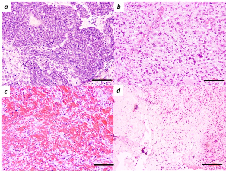
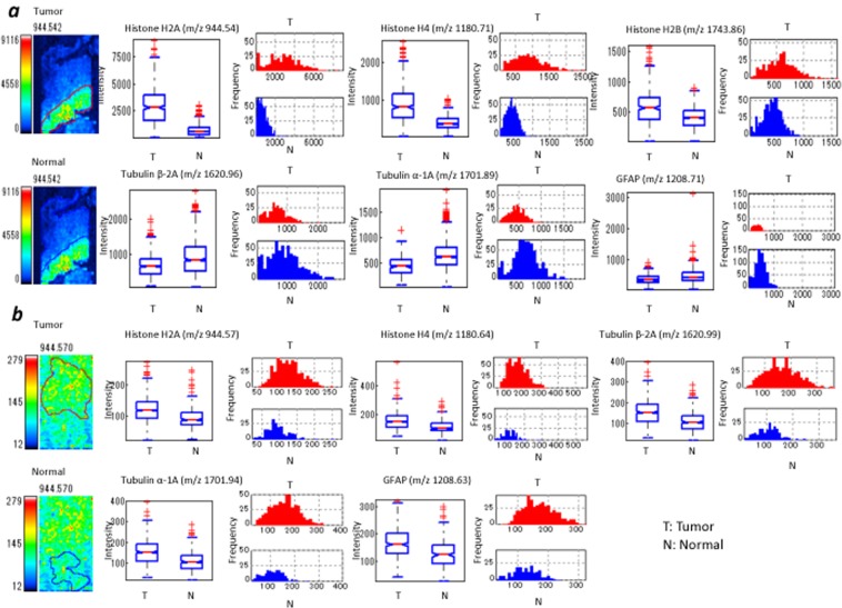
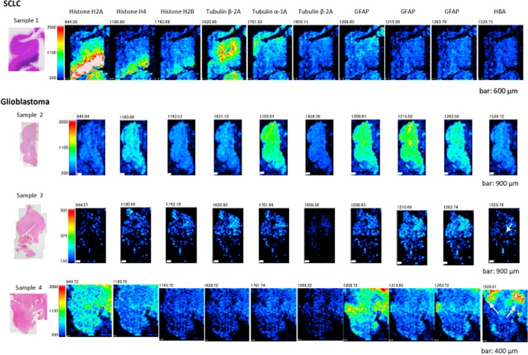
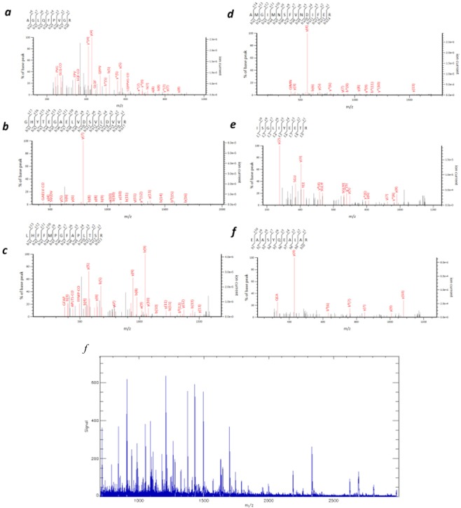
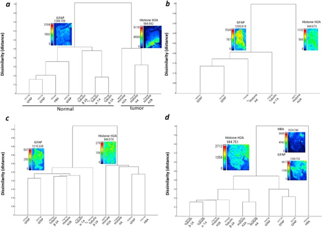
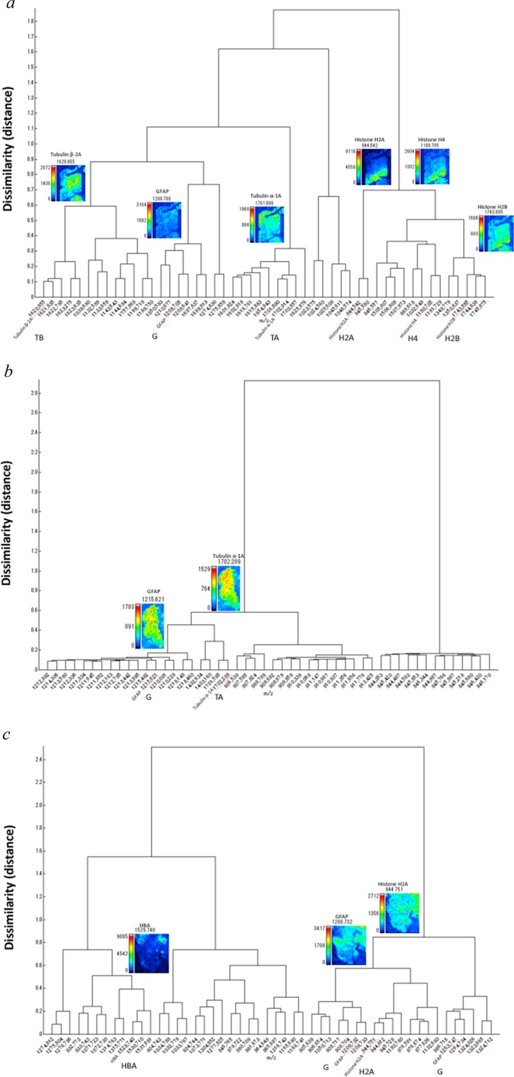
Imaging mass spectrometry (IMS) has been rarely used to examine specimens of human brain tumours. In the current study, high quality brain tumour samples were selected by tissue observation. Further, IMS analysis was combined with a new hierarchical cluster analysis (IMS-HCA) and region of interest analysis (IMS-ROI). IMS-HCA was successful in creating groups consisting of similar signal distribution images of glial fibrillary acidic protein (GFAP) and related multiple proteins in primary brain tumours. This clustering data suggested the relation of GFAP and these identified proteins in the brain tumorigenesis. Also, high levels of histone proteins, haemoglobin subunit α, tubulins, and GFAP were identified in a metastatic brain tumour using IMS-ROI. Our results show that IMS-HCA and IMS-ROI are promising techniques for identifying biomarkers using brain tumour samples.
成像质谱(IMS)很少用于检查人脑肿瘤标本。在本研究中,通过组织观察选择高质量的脑肿瘤样本。此外,IMS 分析与新的层次聚类分析(IMS-HCA)和感兴趣区域分析(IMS-ROI)相结合。IMS-HCA 成功地创建了由原发性脑肿瘤中 GFAP 和相关多种蛋白质的相似信号分布图像组成的组。这些聚类数据表明 GFAP 与脑肿瘤发生过程中的这些鉴定蛋白之间存在关系。此外,使用 IMS-ROI 在转移性脑肿瘤中鉴定到组蛋白、血红蛋白亚基α、微管蛋白和 GFAP 等的高水平。我们的结果表明,IMS-HCA 和 IMS-ROI 是使用脑肿瘤样本鉴定生物标志物的有前途的技术。