• 文献检索
  • 文档翻译
  • 深度研究
  • 学术资讯
  • Suppr Zotero 插件Zotero 插件
  • 邀请有礼
  • 套餐&价格
  • 历史记录
应用&插件
Suppr Zotero 插件Zotero 插件浏览器插件Mac 客户端Windows 客户端微信小程序
定价
高级版会员购买积分包购买API积分包
服务
文献检索文档翻译深度研究API 文档MCP 服务
关于我们
关于 Suppr公司介绍联系我们用户协议隐私条款
关注我们

Suppr 超能文献

核心技术专利:CN118964589B侵权必究
粤ICP备2023148730 号-1Suppr @ 2026

文献检索

告别复杂PubMed语法,用中文像聊天一样搜索,搜遍4000万医学文献。AI智能推荐,让科研检索更轻松。

立即免费搜索

文件翻译

保留排版,准确专业,支持PDF/Word/PPT等文件格式,支持 12+语言互译。

免费翻译文档

深度研究

AI帮你快速写综述,25分钟生成高质量综述,智能提取关键信息,辅助科研写作。

立即免费体验

不同年龄段 COVID-19 感染患者的高分辨率计算机断层扫描表现。

High-resolution computed tomography manifestations of COVID-19 infections in patients of different ages.

机构信息

Department of Radiology, Hangzhou Xixi Hospital Affiliated to Zhejiang Chinese Medical University, Hangzhou, 310023, Zhejiang, China.

Department of Radiology, Sir Run Run Shaw Hospital, School of Medicine, Zhejiang University, Hangzhou, 310023, Zhejiang, China.

出版信息

Eur J Radiol. 2020 May;126:108972. doi: 10.1016/j.ejrad.2020.108972. Epub 2020 Mar 24.

DOI:10.1016/j.ejrad.2020.108972
PMID:32240913
原文链接:https://pmc.ncbi.nlm.nih.gov/articles/PMC7102649/
Abstract

PURPOSE

We aimed to compare chest HRCT lung signs identified in scans of differently aged patients with COVID-19 infections.

METHODS

Case data of patients diagnosed with COVID-19 infection in Hangzhou City, Zhejiang Province in China were collected, and chest HRCT signs of infected patients in four age groups (<18 years, 18-44 years, 45-59 years, ≥60 years) were compared.

RESULTS

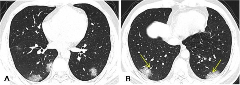
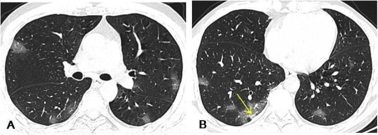
Small patchy, ground-glass opacity (GGO), and consolidations were the main HRCT signs in 98 patients with confirmed COVID-19 infections. Patients aged 45-59 years and aged ≥60 years had more bilateral lung, lung lobe, and lung field involvement, and greater lesion numbers than patients <18 years. GGO accompanied with the interlobular septa thickening or a crazy-paving pattern, consolidation, and air bronchogram sign were more common in patients aged 45-59 years, and ≥60 years, than in those aged <18 years, and aged 18-44 years.

CONCLUSIONS

Chest HRCT manifestations in patients with COVID-19 are related to patient's age, and HRCT signs may be milder in younger patients.

摘要

目的

比较不同年龄段 COVID-19 感染患者的胸部 HRCT 肺部征象。

方法

收集中国浙江省杭州市确诊为 COVID-19 感染患者的病例资料,比较四个年龄组(<18 岁、18-44 岁、45-59 岁、≥60 岁)感染患者的胸部 HRCT 征象。

结果

98 例确诊 COVID-19 感染患者的主要 HRCT 征象为小斑片状、磨玻璃影(GGO)和实变。45-59 岁和≥60 岁年龄组患者双肺、肺叶和肺野受累程度及病变数量均大于<18 岁年龄组。45-59 岁和≥60 岁年龄组患者 GGO 伴间隔增厚或铺路石征、实变和气道征较<18 岁和 18-44 岁年龄组更为常见。

结论

COVID-19 患者的胸部 HRCT 表现与患者年龄有关,年轻患者的 HRCT 征象可能较轻。

https://cdn.ncbi.nlm.nih.gov/pmc/blobs/ce34/7102649/1b88c1456bda/gr3_lrg.jpg
https://cdn.ncbi.nlm.nih.gov/pmc/blobs/ce34/7102649/2ad7e20c3135/gr2_lrg.jpg
https://cdn.ncbi.nlm.nih.gov/pmc/blobs/ce34/7102649/1b88c1456bda/gr3_lrg.jpg
https://cdn.ncbi.nlm.nih.gov/pmc/blobs/ce34/7102649/2ad7e20c3135/gr2_lrg.jpg
https://cdn.ncbi.nlm.nih.gov/pmc/blobs/ce34/7102649/1b88c1456bda/gr3_lrg.jpg

相似文献

1
High-resolution computed tomography manifestations of COVID-19 infections in patients of different ages.不同年龄段 COVID-19 感染患者的高分辨率计算机断层扫描表现。
Eur J Radiol. 2020 May;126:108972. doi: 10.1016/j.ejrad.2020.108972. Epub 2020 Mar 24.
2
Imaging and clinical features of patients with 2019 novel coronavirus SARS-CoV-2.新型冠状病毒 2019 年 SARS-CoV-2 患者的影像学和临床特征。
Eur J Nucl Med Mol Imaging. 2020 May;47(5):1275-1280. doi: 10.1007/s00259-020-04735-9. Epub 2020 Feb 28.
3
Early Clinical and CT Manifestations of Coronavirus Disease 2019 (COVID-19) Pneumonia.新型冠状病毒肺炎(COVID-19)肺炎的早期临床和 CT 表现。
AJR Am J Roentgenol. 2020 Aug;215(2):338-343. doi: 10.2214/AJR.20.22961. Epub 2020 Mar 17.
4
[Clinical features and high resolution CT imaging evolution of coronavirus disease 2019].新型冠状病毒肺炎的临床特征及高分辨率CT影像学演变
Zhonghua Jie He He Hu Xi Za Zhi. 2020 Jun 12;43(6):509-515. doi: 10.3760/cma.j.cn112147-20200214-00094.
5
CT findings of COVID-19 in follow-up: comparison between progression and recovery.新冠肺炎 CT 随访表现:进展与恢复的比较。
Diagn Interv Radiol. 2020 Jul;26(4):301-307. doi: 10.5152/dir.2019.20176.
6
Pulmonary High-Resolution Computed Tomography (HRCT) Findings of Patients with Early-Stage Coronavirus Disease 2019 (COVID-19) in Hangzhou, China.中国杭州早期 2019 冠状病毒病(COVID-19)患者的肺部高分辨率计算机断层扫描(HRCT)表现。
Med Sci Monit. 2020 Apr 4;26:e923885. doi: 10.12659/MSM.923885.
7
CT imaging features of COVID-19 pneumonia: initial experience from Turkey.土耳其的 COVID-19 肺炎 CT 影像学特征:初步经验。
Diagn Interv Radiol. 2020 Jul;26(4):308-314. doi: 10.5152/dir.2020.20307.
8
High-resolution Chest CT Features and Clinical Characteristics of Patients Infected with COVID-19 in Jiangsu, China.中国江苏 COVID-19 感染患者的高分辨率胸部 CT 特征和临床特征。
Int J Infect Dis. 2020 Jun;95:106-112. doi: 10.1016/j.ijid.2020.04.003. Epub 2020 Apr 6.
9
Analysis of clinical features and imaging signs of COVID-19 with the assistance of artificial intelligence.人工智能辅助分析 COVID-19 的临床特征和影像征象。
Eur Rev Med Pharmacol Sci. 2020 Aug;24(15):8210-8218. doi: 10.26355/eurrev_202008_22510.
10
A Comparison of Clinical and Chest CT Findings in Patients With Influenza A (H1N1) Virus Infection and Coronavirus Disease (COVID-19).甲型流感病毒(H1N1)感染与新型冠状病毒肺炎(COVID-19)患者的临床和胸部 CT 表现比较。
AJR Am J Roentgenol. 2020 Nov;215(5):1065-1071. doi: 10.2214/AJR.20.23214. Epub 2020 May 26.

引用本文的文献

1
Factors associated with the intubation of patients with acute respiratory failure and their impact on mortality: a retrospective cohort study.急性呼吸衰竭患者插管相关因素及其对死亡率的影响:一项回顾性队列研究。
BMC Pulm Med. 2025 Jul 3;25(1):308. doi: 10.1186/s12890-025-03773-z.
2
Long-Term Effects of COVID-19: Analysis of Imaging Findings in Patients Evaluated by Computed Tomography from 2020 to 2024.新冠病毒病的长期影响:对2020年至2024年接受计算机断层扫描评估的患者影像结果的分析
Tomography. 2025 Apr 24;11(5):49. doi: 10.3390/tomography11050049.
3
Optimizing Radiation Dose in High-Resolution Chest CT: The Impact of Patient-Specific Factors and Size-Specific Dose Estimates.

本文引用的文献

1
Time Course of Lung Changes at Chest CT during Recovery from Coronavirus Disease 2019 (COVID-19).新冠肺炎(COVID-19)康复过程中胸部 CT 肺部变化的时间进程。
Radiology. 2020 Jun;295(3):715-721. doi: 10.1148/radiol.2020200370. Epub 2020 Feb 13.
2
Diagnosis and treatment recommendations for pediatric respiratory infection caused by the 2019 novel coronavirus.儿童 2019 新型冠状病毒感染所致呼吸道感染的诊断与治疗建议。
World J Pediatr. 2020 Jun;16(3):240-246. doi: 10.1007/s12519-020-00345-5. Epub 2020 Feb 5.
3
CT Imaging Features of 2019 Novel Coronavirus (2019-nCoV).
优化高分辨率胸部CT的辐射剂量:患者特定因素和尺寸特定剂量估计的影响
Diagnostics (Basel). 2025 Mar 16;15(6):740. doi: 10.3390/diagnostics15060740.
4
Association of chest computed tomography severity score at ICU admission and respiratory outcomes in critically ill COVID-19 patients.入住 ICU 时的胸部 CT 严重程度评分与危重症 COVID-19 患者的呼吸结局的相关性。
PLoS One. 2024 May 2;19(5):e0299390. doi: 10.1371/journal.pone.0299390. eCollection 2024.
5
Lung abnormalities on computed tomography of Vietnamese patients with COVID-19 and the association with medical variables.越南新冠肺炎患者计算机断层扫描的肺部异常情况及其与医学变量的关联。
IJID Reg. 2024 Jan 19;10:183-190. doi: 10.1016/j.ijregi.2024.01.006. eCollection 2024 Mar.
6
Comparison of Chest Computed Tomography (CT) Imaging Patterns and Severity among COVID-19 Patients during the First and Fourth Waves in Addis Ababa, Ethiopia.埃塞俄比亚亚的斯亚贝巴新冠疫情第一波和第四波期间新冠肺炎患者胸部计算机断层扫描(CT)成像模式与严重程度的比较
Radiol Res Pract. 2023 Nov 29;2023:6385162. doi: 10.1155/2023/6385162. eCollection 2023.
7
Investigating of the role of CT scan for cancer patients during the first wave of COVID-19 pandemic.在新冠疫情第一波期间对癌症患者进行CT扫描作用的调查。
Res Diagn Interv Imaging. 2022 Mar;1:100004. doi: 10.1016/j.redii.2022.100004. Epub 2022 Mar 31.
8
SARS-CoV-2 Infection and COVID-19 in Children.儿童感染 SARS-CoV-2 病毒和 COVID-19 疾病。
Clin Chest Med. 2023 Jun;44(2):359-371. doi: 10.1016/j.ccm.2022.11.014. Epub 2022 Nov 22.
9
Novel Corona Virus 2019 Disease: Assessment on High-Resolution Computed Tomography Thorax.2019新型冠状病毒病:胸部高分辨率计算机断层扫描评估
Cureus. 2023 Feb 26;15(2):e35506. doi: 10.7759/cureus.35506. eCollection 2023 Feb.
10
CT Examinations for COVID-19: A Systematic Review of Protocols, Radiation Dose, and Numbers Needed to Diagnose and Predict.用于新型冠状病毒肺炎的CT检查:对方案、辐射剂量以及诊断和预测所需数量的系统评价
Taehan Yongsang Uihakhoe Chi. 2021 Nov;82(6):1505-1523. doi: 10.3348/jksr.2021.0096. Epub 2021 Nov 4.
2019 新型冠状病毒(2019-nCoV)的 CT 影像学特征。
Radiology. 2020 Apr;295(1):202-207. doi: 10.1148/radiol.2020200230. Epub 2020 Feb 4.
4
A Novel Coronavirus from Patients with Pneumonia in China, 2019.2019 年中国肺炎患者中的一种新型冠状病毒。
N Engl J Med. 2020 Feb 20;382(8):727-733. doi: 10.1056/NEJMoa2001017. Epub 2020 Jan 24.
5
Radiographic and CT Features of Viral Pneumonia.病毒性肺炎的影像学和 CT 特征。
Radiographics. 2018 May-Jun;38(3):719-739. doi: 10.1148/rg.2018170048.
6
Radio-pathological correlation of organizing pneumonia (OP): a pictorial review.机化性肺炎(OP)的放射学与病理学相关性:图文综述
Br J Radiol. 2017 Mar;90(1071):20160723. doi: 10.1259/bjr.20160723. Epub 2017 Feb 17.