• 文献检索
  • 文档翻译
  • 深度研究
  • 学术资讯
  • Suppr Zotero 插件Zotero 插件
  • 邀请有礼
  • 套餐&价格
  • 历史记录
应用&插件
Suppr Zotero 插件Zotero 插件浏览器插件Mac 客户端Windows 客户端微信小程序
定价
高级版会员购买积分包购买API积分包
服务
文献检索文档翻译深度研究API 文档MCP 服务
关于我们
关于 Suppr公司介绍联系我们用户协议隐私条款
关注我们

Suppr 超能文献

核心技术专利:CN118964589B侵权必究
粤ICP备2023148730 号-1Suppr @ 2026

文献检索

告别复杂PubMed语法,用中文像聊天一样搜索,搜遍4000万医学文献。AI智能推荐,让科研检索更轻松。

立即免费搜索

文件翻译

保留排版,准确专业,支持PDF/Word/PPT等文件格式,支持 12+语言互译。

免费翻译文档

深度研究

AI帮你快速写综述,25分钟生成高质量综述,智能提取关键信息,辅助科研写作。

立即免费体验

新型冠状病毒 2019 年 SARS-CoV-2 患者的影像学和临床特征。

Imaging and clinical features of patients with 2019 novel coronavirus SARS-CoV-2.

机构信息

Department of Medical Imaging Center, The First Affiliated Hospital, Jinan University, No. 613, Huangpu Road West, Tianhe District, Guangzhou, 510630, Guangdong Province, China.

Department of Radiology, Guangzhou Eighth People's Hospital, Guangzhou Medical University, No. 8, Huaying Road, Baiyun District, Guangzhou, 510060, Guangdong Province, China.

出版信息

Eur J Nucl Med Mol Imaging. 2020 May;47(5):1275-1280. doi: 10.1007/s00259-020-04735-9. Epub 2020 Feb 28.

DOI:10.1007/s00259-020-04735-9
PMID:32107577
原文链接:https://pmc.ncbi.nlm.nih.gov/articles/PMC7080117/
Abstract

BACKGROUND

The pneumonia caused by the 2019 novel coronavirus (SARS-CoV-2, also called 2019-nCoV) recently break out in Wuhan, China, and was named as COVID-19. With the spread of the disease, similar cases have also been confirmed in other regions of China. We aimed to report the imaging and clinical characteristics of these patients infected with SARS-CoV-2 in Guangzhou, China.

METHODS

All patients with laboratory-identified SARS-CoV-2 infection by real-time polymerase chain reaction (PCR) were collected between January 23, 2020, and February 4, 2020, in a designated hospital (Guangzhou Eighth People's Hospital). This analysis included 90 patients (39 men and 51 women; median age, 50 years (age range, 18-86 years). All the included SARS-CoV-2-infected patients underwent non-contrast enhanced chest computed tomography (CT). We analyzed the clinical characteristics of the patients, as well as the distribution characteristics, pattern, morphology, and accompanying manifestations of lung lesions. In addition, after 1-6 days (mean 3.5 days), follow-up chest CT images were evaluated to assess radiological evolution.

FINDINGS

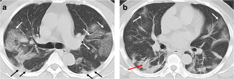
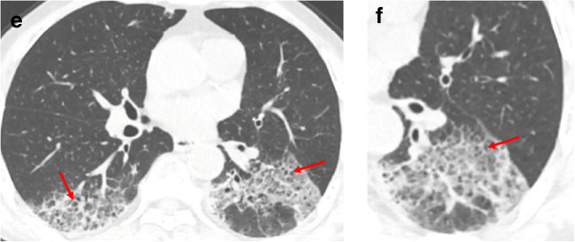
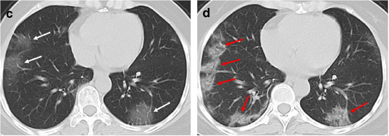
The majority of infected patients had a history of exposure in Wuhan or to infected patients and mostly presented with fever and cough. More than half of the patients presented bilateral, multifocal lung lesions, with peripheral distribution, and 53 (59%) patients had more than two lobes involved. Of all included patients, COVID-19 pneumonia presented with ground glass opacities in 65 (72%), consolidation in 12 (13%), crazy paving pattern in 11 (12%), interlobular thickening in 33 (37%), adjacent pleura thickening in 50 (56%), and linear opacities combined in 55 (61%). Pleural effusion, pericardial effusion, and lymphadenopathy were uncommon findings. In addition, baseline chest CT did not show any abnormalities in 21 patients (23%), but 3 patients presented bilateral ground glass opacities on the second CT after 3-4 days.

CONCLUSION

SARS-CoV-2 infection can be confirmed based on the patient's history, clinical manifestations, imaging characteristics, and laboratory tests. Chest CT examination plays an important role in the initial diagnosis of the novel coronavirus pneumonia. Multiple patchy ground glass opacities in bilateral multiple lobular with periphery distribution are typical chest CT imaging features of the COVID-19 pneumonia.

摘要

背景

近期在中国武汉爆发的由 2019 年新型冠状病毒(SARS-CoV-2,也称为 2019-nCoV)引起的肺炎,被命名为 COVID-19。随着疾病的传播,中国其他地区也确诊了类似病例。我们旨在报告在中国广州感染 SARS-CoV-2 的这些患者的影像学和临床特征。

方法

2020 年 1 月 23 日至 2 月 4 日,我们在一家指定医院(广州第八人民医院)收集了所有经实时聚合酶链反应(PCR)检测证实的 SARS-CoV-2 感染患者。本分析包括 90 例患者(39 名男性和 51 名女性;中位年龄 50 岁(年龄范围 18-86 岁)。所有纳入的 SARS-CoV-2 感染患者均接受非增强胸部 CT 检查。我们分析了患者的临床特征,以及肺部病变的分布特征、模式、形态和伴随表现。此外,在 1-6 天后(平均 3.5 天),评估随访胸部 CT 图像以评估影像学演变。

结果

大多数感染患者有武汉接触史或感染患者接触史,主要表现为发热和咳嗽。超过一半的患者表现为双侧、多灶性肺部病变,呈外周分布,53 例(59%)患者有两个以上肺叶受累。所有纳入患者中,COVID-19 肺炎 65 例(72%)表现为磨玻璃影,12 例(13%)为实变,11 例(12%)为铺路石征,33 例(37%)为小叶间隔增厚,50 例(56%)为邻近胸膜增厚,55 例(61%)为线状混浊合并。胸腔积液、心包积液和淋巴结病少见。此外,21 例患者(23%)基线胸部 CT 无任何异常,但 3 例患者在第 3-4 天后第二次 CT 检查时出现双侧磨玻璃影。

结论

根据患者的病史、临床表现、影像学特征和实验室检查可以确诊 SARS-CoV-2 感染。胸部 CT 检查在新型冠状病毒肺炎的初步诊断中起重要作用。双侧多发性肺叶多灶性分布的多发性斑片状磨玻璃影是 COVID-19 肺炎的典型胸部 CT 影像学特征。

https://cdn.ncbi.nlm.nih.gov/pmc/blobs/bc0a/7080117/1a55b08b6105/259_2020_4735_Fig3_HTML.jpg
https://cdn.ncbi.nlm.nih.gov/pmc/blobs/bc0a/7080117/2d8dc701dbf2/259_2020_4735_Fig1_HTML.jpg
https://cdn.ncbi.nlm.nih.gov/pmc/blobs/bc0a/7080117/155a0af74c8a/259_2020_4735_Fig2_HTML.jpg
https://cdn.ncbi.nlm.nih.gov/pmc/blobs/bc0a/7080117/1a55b08b6105/259_2020_4735_Fig3_HTML.jpg
https://cdn.ncbi.nlm.nih.gov/pmc/blobs/bc0a/7080117/2d8dc701dbf2/259_2020_4735_Fig1_HTML.jpg
https://cdn.ncbi.nlm.nih.gov/pmc/blobs/bc0a/7080117/155a0af74c8a/259_2020_4735_Fig2_HTML.jpg
https://cdn.ncbi.nlm.nih.gov/pmc/blobs/bc0a/7080117/1a55b08b6105/259_2020_4735_Fig3_HTML.jpg

相似文献

1
Imaging and clinical features of patients with 2019 novel coronavirus SARS-CoV-2.新型冠状病毒 2019 年 SARS-CoV-2 患者的影像学和临床特征。
Eur J Nucl Med Mol Imaging. 2020 May;47(5):1275-1280. doi: 10.1007/s00259-020-04735-9. Epub 2020 Feb 28.
2
CT imaging changes of corona virus disease 2019(COVID-19): a multi-center study in Southwest China.COVID-19 的 CT 影像学改变:中国西南部多中心研究。
J Transl Med. 2020 Apr 6;18(1):154. doi: 10.1186/s12967-020-02324-w.
3
A Comparison of Clinical and Chest CT Findings in Patients With Influenza A (H1N1) Virus Infection and Coronavirus Disease (COVID-19).甲型流感病毒(H1N1)感染与新型冠状病毒肺炎(COVID-19)患者的临床和胸部 CT 表现比较。
AJR Am J Roentgenol. 2020 Nov;215(5):1065-1071. doi: 10.2214/AJR.20.23214. Epub 2020 May 26.
4
Emerging 2019 Novel Coronavirus (2019-nCoV) Pneumonia.新型冠状病毒(2019-nCoV)肺炎的诊治进展。
Radiology. 2020 Apr;295(1):210-217. doi: 10.1148/radiol.2020200274. Epub 2020 Feb 6.
5
Chest CT imaging characteristics of COVID-19 pneumonia in preschool children: a retrospective study.儿童 COVID-19 肺炎的胸部 CT 影像学特征:一项回顾性研究。
BMC Pediatr. 2020 May 18;20(1):227. doi: 10.1186/s12887-020-02140-7.
6
Clinical and computed tomographic imaging features of novel coronavirus pneumonia caused by SARS-CoV-2.SARS-CoV-2 引起的新型冠状病毒肺炎的临床和计算机断层扫描影像学特征。
J Infect. 2020 Apr;80(4):394-400. doi: 10.1016/j.jinf.2020.02.017. Epub 2020 Feb 25.
7
Dynamic Chest CT Evaluation in Three Cases of 2019 Novel Coronavirus Pneumonia.动态胸部 CT 评估 2019 年新型冠状病毒肺炎三例
Arch Iran Med. 2020 Apr 1;23(4):277-280. doi: 10.34172/aim.2020.11.
8
Clinical Features and Chest CT Manifestations of Coronavirus Disease 2019 (COVID-19) in a Single-Center Study in Shanghai, China.中国上海单中心研究的 2019 年冠状病毒病(COVID-19)的临床特征和胸部 CT 表现。
AJR Am J Roentgenol. 2020 Jul;215(1):121-126. doi: 10.2214/AJR.20.22959. Epub 2020 Mar 14.
9
[Preliminary Study on Clinical Features and CT Findings of Common-type Coronavirus Disease 2019 Patients in Peking Union Medical College Hospital].北京协和医院2019新型冠状病毒病普通型患者临床特征及CT表现的初步研究
Zhongguo Yi Xue Ke Xue Yuan Xue Bao. 2020 Jun 30;42(3):376-382. doi: 10.3881/j.issn.1000-503X.12588.
10
[Analysis of CT features of 15 children with 2019 novel coronavirus infection].[15例2019新型冠状病毒感染儿童的CT特征分析]
Zhonghua Er Ke Za Zhi. 2020 Apr 2;58(4):275-278. doi: 10.3760/cma.j.cn112140-20200210-00071.

引用本文的文献

1
Prevalence of comorbidities and their association with disease severity and mortality in COVID-19 patients: A systematic review and meta-analysis.COVID-19患者合并症的患病率及其与疾病严重程度和死亡率的关联:一项系统综述和荟萃分析。
J Multimorb Comorb. 2025 Aug 28;15:26335565251371256. doi: 10.1177/26335565251371256. eCollection 2025 Jan-Dec.
2
Artificial intelligence algorithms based approach in evaluating COVID-19 patients and management.基于人工智能算法的方法在评估新冠肺炎患者及管理中的应用。
J Crit Care Med (Targu Mures). 2025 Jul 31;11(3):247-256. doi: 10.2478/jccm-2025-0032. eCollection 2025 Jul.
3

本文引用的文献

1
CT Imaging Features of 2019 Novel Coronavirus (2019-nCoV).2019 新型冠状病毒(2019-nCoV)的 CT 影像学特征。
Radiology. 2020 Apr;295(1):202-207. doi: 10.1148/radiol.2020200230. Epub 2020 Feb 4.
2
Evolution of the novel coronavirus from the ongoing Wuhan outbreak and modeling of its spike protein for risk of human transmission.新型冠状病毒在武汉持续爆发中的进化及其刺突蛋白对人类传播风险的建模。
Sci China Life Sci. 2020 Mar;63(3):457-460. doi: 10.1007/s11427-020-1637-5. Epub 2020 Jan 21.
3
Epidemiological and clinical characteristics of 99 cases of 2019 novel coronavirus pneumonia in Wuhan, China: a descriptive study.
Integrating CT radiomics and clinical features using machine learning to predict post-COVID pulmonary fibrosis.
利用机器学习整合CT影像组学和临床特征以预测新冠后肺纤维化
Respir Res. 2025 Jul 2;26(1):227. doi: 10.1186/s12931-025-03305-7.
4
Assessment of upper respiratory and gut bacterial microbiomes during COVID-19 infection in adults: potential aerodigestive transmission.成人新冠病毒感染期间上呼吸道和肠道细菌微生物群评估:气消化道传播的可能性
Sci Rep. 2025 Jan 13;15(1):1811. doi: 10.1038/s41598-025-85806-5.
5
The influence of Omicron on vaccine efficacy and durability: a neurology perspective.奥密克戎对疫苗效力和持久性的影响:神经学视角
Clin Exp Vaccine Res. 2024 Jul;13(3):175-183. doi: 10.7774/cevr.2024.13.3.175. Epub 2024 Jul 31.
6
Pulmonary microbial spectrum in late-stage SARS-CoV-2 infection: a case series.晚期 SARS-CoV-2 感染的肺部微生物谱:病例系列。
Eur J Clin Microbiol Infect Dis. 2024 Oct;43(10):2037-2046. doi: 10.1007/s10096-024-04897-6. Epub 2024 Jul 20.
7
Community-Based View on Diagnostic Imaging at the End of COVID-19 Pandemic: Online Survey-Assisted Study.新冠疫情末期基于社区的诊断成像观点:在线调查辅助研究
Diagnostics (Basel). 2024 Jun 15;14(12):1269. doi: 10.3390/diagnostics14121269.
8
Targeting GPVI with glenzocimab in COVID-19 patients: Results from a randomized clinical trial.以 glenzocimab 靶向 GPVI 在 COVID-19 患者中的应用:一项随机临床试验结果。
PLoS One. 2024 Jun 17;19(6):e0302897. doi: 10.1371/journal.pone.0302897. eCollection 2024.
9
A Retrospective Study of the Epidemiologic and Clinical Characteristics of COVID-19 Among Hospitalized Patients in Quanzhou, China.中国泉州住院患者中 COVID-19 的流行病学和临床特征的回顾性研究
Infect Microbes Dis. 2021 Jan 15;3(1):32-40. doi: 10.1097/IM9.0000000000000048. eCollection 2021 Mar.
10
[Intensive care during the 2019-coronavirus epidemic].[2019冠状病毒疫情期间的重症监护]
Med Intensiva. 2020 Aug-Sep;44(6):351-362. doi: 10.1016/j.medin.2020.03.001. Epub 2020 Mar 30.
中国武汉 99 例 2019 年新型冠状病毒肺炎患者的流行病学和临床特征:描述性研究。
Lancet. 2020 Feb 15;395(10223):507-513. doi: 10.1016/S0140-6736(20)30211-7. Epub 2020 Jan 30.
4
Clinical features of patients infected with 2019 novel coronavirus in Wuhan, China.中国武汉地区 2019 年新型冠状病毒感染患者的临床特征。
Lancet. 2020 Feb 15;395(10223):497-506. doi: 10.1016/S0140-6736(20)30183-5. Epub 2020 Jan 24.
5
A familial cluster of pneumonia associated with the 2019 novel coronavirus indicating person-to-person transmission: a study of a family cluster.一个涉及 2019 年新型冠状病毒的家庭聚集性肺炎病例,提示存在人际传播:一项家庭聚集性研究。
Lancet. 2020 Feb 15;395(10223):514-523. doi: 10.1016/S0140-6736(20)30154-9. Epub 2020 Jan 24.
6
Data sharing and outbreaks: best practice exemplified.数据共享与疫情爆发:最佳实践示例
Lancet. 2020 Feb 15;395(10223):469-470. doi: 10.1016/S0140-6736(20)30184-7. Epub 2020 Jan 24.
7
A Novel Coronavirus from Patients with Pneumonia in China, 2019.2019 年中国肺炎患者中的一种新型冠状病毒。
N Engl J Med. 2020 Feb 20;382(8):727-733. doi: 10.1056/NEJMoa2001017. Epub 2020 Jan 24.
8
Origin and evolution of pathogenic coronaviruses.致病冠状病毒的起源与演化。
Nat Rev Microbiol. 2019 Mar;17(3):181-192. doi: 10.1038/s41579-018-0118-9.
9
Radiographic and CT Features of Viral Pneumonia.病毒性肺炎的影像学和 CT 特征。
Radiographics. 2018 May-Jun;38(3):719-739. doi: 10.1148/rg.2018170048.
10
Epidemiology, Genetic Recombination, and Pathogenesis of Coronaviruses.冠状病毒的流行病学、基因重组与发病机制
Trends Microbiol. 2016 Jun;24(6):490-502. doi: 10.1016/j.tim.2016.03.003. Epub 2016 Mar 21.