Lipodystrophies, Metabolic and Hormonal Adaptation, and Aging, UMR_S 938, Centre de Recherche Saint-Antoine-Institut Hospitalo-Universitaire de Cardiométabolisme et Nutrition (ICAN), INSERM, Sorbonne Université, F-75012 Paris, France.
Immunology of Viral infections and Autoimmune Diseases, IDMIT Department, IBFJ, U1184, INSERM -CEA-Université Paris Sud 11, F-92260 Fontenay-Aux-Roses and F-94270 Le Kremlin-Bicêtre, France.
Cells. 2020 Apr 1;9(4):854. doi: 10.3390/cells9040854.
Aging is characterized by adipose tissue senescence, inflammation, and fibrosis, with trunk fat accumulation. Aging HIV-infected patients have a higher risk of trunk fat accumulation than uninfected individuals-suggesting that viral infection has a role in adipose tissue aging. We previously demonstrated that HIV/SIV infection and the Tat and Nef viral proteins were responsible for adipose tissue fibrosis and impaired adipogenesis. We hypothesized that SIV/HIV infection and viral proteins could induce adipose tissue senescence and thus lead to adipocyte dysfunctions.
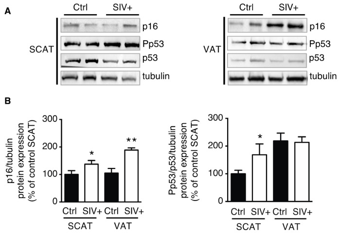
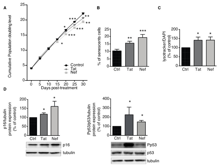
Features of tissue senescence were evaluated in subcutaneous and visceral adipose tissues of SIV-infected macaques and in human adipose stem cells (ASCs) exposed to Tat or Nef for up to 30 days.
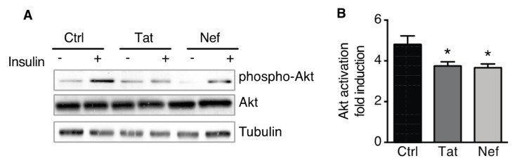
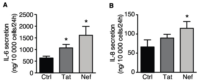
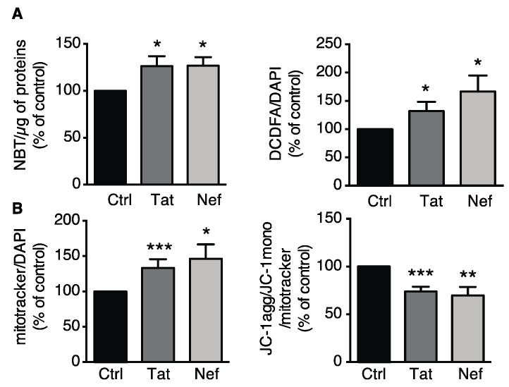
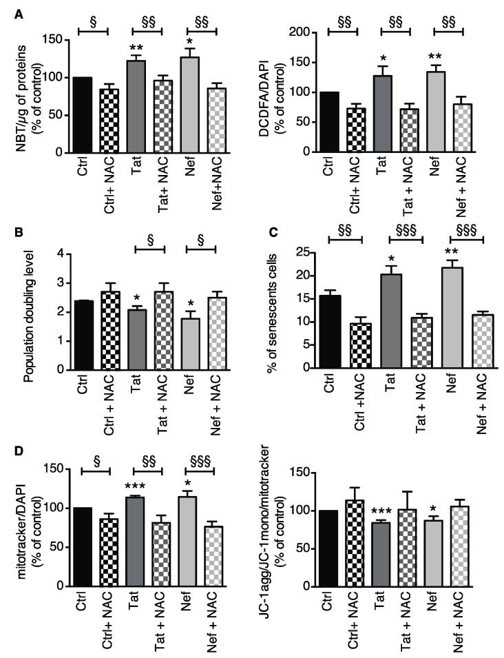
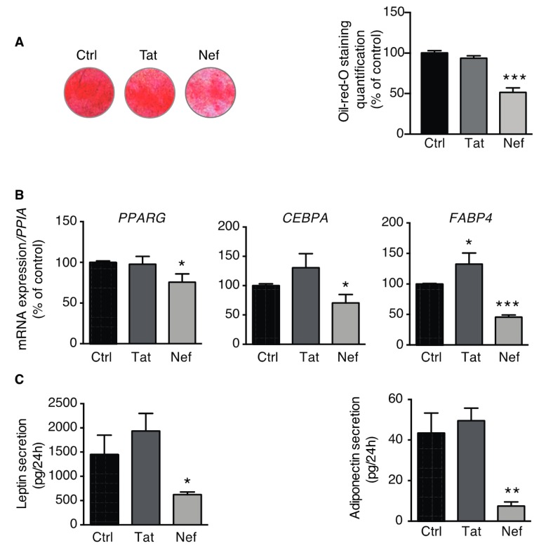
p16 expression and p53 activation were higher in adipose tissue of SIV-infected macaques than in control macaques, indicating adipose tissue senescence. Tat and Nef induced higher senescence in ASCs, characterized by higher levels of senescence-associated beta-galactosidase activity, p16 expression, and p53 activation vs. control cells. Treatment with Tat and Nef also induced oxidative stress and mitochondrial dysfunction. Prevention of oxidative stress (using N-acetyl-cysteine) reduced senescence in ASCs. Adipocytes having differentiated from Nef-treated ASCs displayed alterations in adipogenesis with lower levels of triglyceride accumulation and adipocyte marker expression and secretion, and insulin resistance.
HIV/SIV promotes adipose tissue senescence, which in turn may alter adipocyte function and contribute to insulin resistance.
衰老的特征是脂肪组织衰老、炎症和纤维化,以及躯干脂肪堆积。与未感染个体相比,衰老的 HIV 感染者更容易出现躯干脂肪堆积,这表明病毒感染在脂肪组织衰老中起作用。我们之前的研究表明,HIV/SIV 感染以及 Tat 和 Nef 病毒蛋白与脂肪组织纤维化和脂肪生成受损有关。我们假设 SIV/HIV 感染和病毒蛋白可诱导脂肪组织衰老,从而导致脂肪细胞功能障碍。
在 SIV 感染的猕猴的皮下和内脏脂肪组织中以及在暴露于 Tat 或 Nef 长达 30 天的人脂肪干细胞 (ASC) 中评估组织衰老的特征。
与对照猕猴相比,SIV 感染猕猴的脂肪组织中 p16 表达和 p53 激活增加,表明脂肪组织衰老。与对照细胞相比,Tat 和 Nef 诱导 ASC 中更高的衰老,其特征为衰老相关的β-半乳糖苷酶活性、p16 表达和 p53 激活水平更高。用 Tat 和 Nef 处理还诱导氧化应激和线粒体功能障碍。用 N-乙酰半胱氨酸预防氧化应激可减少 ASC 中的衰老。用 Nef 处理的 ASC 分化而来的脂肪细胞在脂肪生成方面表现出改变,其甘油三酯积累、脂肪细胞标志物表达和分泌以及胰岛素抵抗水平降低。
HIV/SIV 促进脂肪组织衰老,进而可能改变脂肪细胞功能并导致胰岛素抵抗。