Department of Environmental Sciences and Engineering, Government College University, Faisalabad, Pakistan.
National Centre of Excellence in Molecular Biology, University of the Punjab, Lahore, Pakistan.
Sci Rep. 2020 Apr 3;10(1):5897. doi: 10.1038/s41598-020-62807-0.
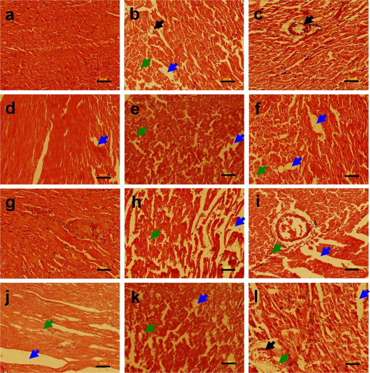
Diabetes is a complex metabolic disorder and different environmental toxicants including heavy metals have been involved in diabetes induction. Therefore, assessment of the environmental risk factors and heavy metals induced toxicity have become critical for reducing the consequences of metals pollutants. Previously, we reported heavy metals induced nephrotoxicity in non-diabetic and diabetic rats. Here, we extended our analysis by examining the heavy metals induced organs (heart, kidney, liver, pancreas, and spleen) damage in diabetic and non-diabetic Wistar rats using histopathology and quantitative real-time PCR (qRT-PCR). Following the generation of the diabetic rat model, the animals were exposed to heavy metals including lead (Pb), arsenic (As), manganese (Mn) and cadmium (Cd). Both non-diabetic and diabetic rats were exposed to heavy metals for 30 days and subsequently, the heart, kidney, liver, pancreas and spleen tissues were examined. Heavy metal treatment resulted in irregularly arranged myofibrils and vacuolization in the heart tissue of metal treated groups as evident from hematoxylin and eosin (H & E) staining. The kidney tissue of rats treated with heavy metals showed tubular degeneration, fibrosis, hemorrhage, and vacuolation. The liver of the heavy metals treated rats exhibited cellular degeneration and necrosis. The pancreatic tissue of streptozotocin injected untreated and metal treated rats revealed severe degeneration, necrosis, degranulation, shrinkage, and depression in the islets of Langerhans. Increased red pulp area and congestion were observed in the spleen of the metal mixture treated non-diabetic and diabetic rats. In line with the histological data, the qRT-PCR analysis showed downregulated expression of Bcl and upregulation of Caspase-3 in non-diabetic and diabetic metal treated rats as compared to the non-diabetic untreated rats. In conclusion, the present study revealed, diabetic rats are more prone to metal alone as well as metal mixture induced organ damage as compared to non-diabetic rats.
糖尿病是一种复杂的代谢紊乱疾病,包括重金属在内的不同环境毒物已被涉及到糖尿病的诱导中。因此,评估环境风险因素和重金属诱导的毒性对于减少金属污染物的后果变得至关重要。此前,我们报道了重金属在非糖尿病和糖尿病大鼠中诱导的肾毒性。在这里,我们通过使用组织病理学和实时定量 PCR(qRT-PCR)检查糖尿病和非糖尿病 Wistar 大鼠中重金属诱导的器官(心脏、肾脏、肝脏、胰腺和脾脏)损伤,扩展了我们的分析。在生成糖尿病大鼠模型后,将动物暴露于包括铅(Pb)、砷(As)、锰(Mn)和镉(Cd)在内的重金属中。非糖尿病和糖尿病大鼠均暴露于重金属 30 天,随后检查心脏、肾脏、肝脏、胰腺和脾脏组织。重金属处理导致金属处理组的心肌中肌原纤维排列不规则和空泡化,如苏木精和伊红(H&E)染色所示。重金属处理大鼠的肾脏组织显示管状变性、纤维化、出血和空泡化。重金属处理大鼠的肝脏表现出细胞变性和坏死。链脲佐菌素注射的未处理和金属处理大鼠的胰腺组织显示严重的变性、坏死、脱颗粒、萎缩和胰岛兰格汉斯岛的凹陷。金属混合物处理的非糖尿病和糖尿病大鼠的脾脏中观察到红髓面积增加和充血。与组织学数据一致,qRT-PCR 分析显示,与非糖尿病未处理大鼠相比,非糖尿病和糖尿病金属处理大鼠的 Bcl 表达下调,Caspase-3 表达上调。总之,本研究表明,与非糖尿病大鼠相比,糖尿病大鼠更容易受到单一金属以及金属混合物诱导的器官损伤。