TBI, CNRS, INRAE, INSA, Université de Toulouse, Toulouse, France.
Laboratoire des Venins et Molécules Thérapeutiques, Institut Pasteur de Tunis, Université Tunis El Manar, Tunis, Tunisia.
Microbiologyopen. 2020 Jun;9(6):1175-1182. doi: 10.1002/mbo3.1027. Epub 2020 Apr 7.
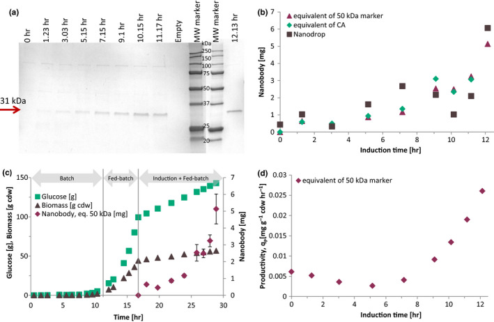
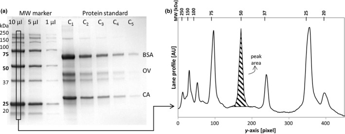
The protein purity is generally checked using SDS-PAGE, where densitometry could be used to quantify the protein bands. In literature, few studies have been reported using image analysis for the quantification of protein in SDS-PAGE: that is, imaged with Stain-Free™ technology. This study presents a protocol of image analysis for electrophoresis gels that allows the quantification of unknown proteins using the molecular weight markers as protein standards. Escherichia coli WK6/pHEN6 encoding the bispecific nanobody CH10-12 engineered by the Pasteur Institute of Tunisia was cultured in a bioreactor and induced with isopropyl β-D-1-thiogalactopyranoside (IPTG) at 28°C for 12 hr. Periplasmic proteins extracted by osmotic shock were purified by immobilized metal affinity chromatography (IMAC). Images of the SDS-PAGE gels were analyzed using ImageJ, and the lane profiles were obtained in grayscale and uncalibrated optical density. Protein load and peak area were linearly correlated, and optimal image processing was then performed by background subtraction using the rolling ball algorithm with radius size 250 pixels. No brightness and contrast adjustment was applied. The production of the nanobody CH10-12 was obtained through a fed-batch strategy and quantified using the band of 50 kDa in the marker as reference for 750 ng of recombinant protein. The molecular weight marker was used as a sole protein standard for protein quantification in SDS-PAGE gel images.
蛋白质纯度通常使用 SDS-PAGE 进行检查,其中密度测定法可用于定量蛋白质条带。在文献中,很少有研究使用图像分析来定量 SDS-PAGE 中的蛋白质:即使用 Stain-Free™ 技术进行成像。本研究提出了一种电泳凝胶的图像分析方案,允许使用分子量标志物作为蛋白质标准来定量未知蛋白质。由突尼斯巴斯德研究所设计的双特异性纳米抗体 CH10-12 的大肠杆菌 WK6/pHEN6 在生物反应器中培养,并在 28°C 下用异丙基 β-D-1-硫代半乳糖吡喃糖苷 (IPTG) 诱导 12 小时。通过渗透压休克提取的周质蛋白通过固定化金属亲和层析 (IMAC) 进行纯化。使用 ImageJ 分析 SDS-PAGE 凝胶的图像,并以灰度和未经校准的光密度获得泳道轮廓。蛋白质负载和峰面积呈线性相关,然后通过使用半径大小为 250 像素的滚动球算法进行背景扣除来进行最佳图像处理。未应用亮度和对比度调整。纳米抗体 CH10-12 的生产通过分批补料策略获得,并使用标记物中的 50 kDa 条带作为参考,对 750 ng 重组蛋白进行定量。分子量标志物被用作 SDS-PAGE 凝胶图像中蛋白质定量的唯一蛋白质标准。