• 文献检索
  • 文档翻译
  • 深度研究
  • 学术资讯
  • Suppr Zotero 插件Zotero 插件
  • 邀请有礼
  • 套餐&价格
  • 历史记录
应用&插件
Suppr Zotero 插件Zotero 插件浏览器插件Mac 客户端Windows 客户端微信小程序
定价
高级版会员购买积分包购买API积分包
服务
文献检索文档翻译深度研究API 文档MCP 服务
关于我们
关于 Suppr公司介绍联系我们用户协议隐私条款
关注我们

Suppr 超能文献

核心技术专利:CN118964589B侵权必究
粤ICP备2023148730 号-1Suppr @ 2026

文献检索

告别复杂PubMed语法,用中文像聊天一样搜索,搜遍4000万医学文献。AI智能推荐,让科研检索更轻松。

立即免费搜索

文件翻译

保留排版,准确专业,支持PDF/Word/PPT等文件格式,支持 12+语言互译。

免费翻译文档

深度研究

AI帮你快速写综述,25分钟生成高质量综述,智能提取关键信息,辅助科研写作。

立即免费体验

荧光光漂白作为定量扩展显微镜中生物样品三维扩展因子的内在工具。

Fluorescence Photobleaching as an Intrinsic Tool to Quantify the 3D Expansion Factor of Biological Samples in Expansion Microscopy.

作者信息

Vanheusden Marisa, Vitale Raffaele, Camacho Rafael, Janssen Kris P F, Acke Aline, Rocha Susana, Hofkens Johan

机构信息

Department of Chemistry, KU Leuven, Leuven 3000, Belgium.

出版信息

ACS Omega. 2020 Mar 17;5(12):6792-6799. doi: 10.1021/acsomega.0c00118. eCollection 2020 Mar 31.

DOI:10.1021/acsomega.0c00118
PMID:32258914
原文链接:https://pmc.ncbi.nlm.nih.gov/articles/PMC7114699/
Abstract

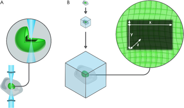
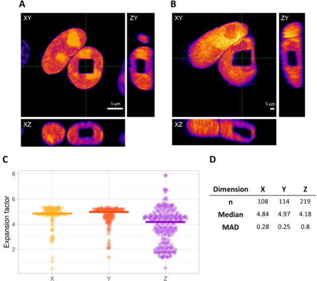
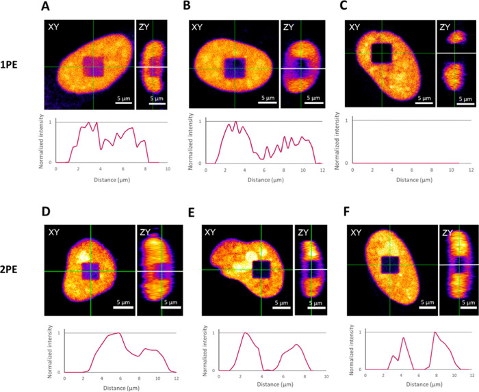
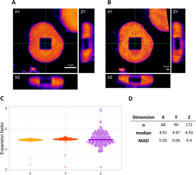
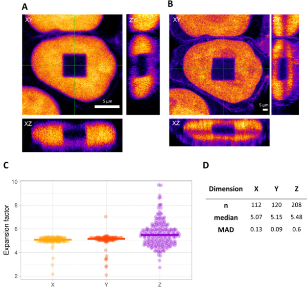
Four years after its first report, expansion microscopy (ExM) is now being routinely applied in laboratories worldwide to achieve super-resolution imaging on conventional fluorescence microscopes. By chemically anchoring all molecules of interest to the polymer meshwork of an expandable hydrogel, their physical distance is increased by a factor of ∼4-5× upon dialysis in water, resulting in an imprint of the original sample with a lateral resolution up to 50-70 nm. To ensure a correct representation of the original spatial distribution of the molecules, it is crucial to confirm that the expansion is isotropic, preferentially in all three dimensions. To address this, we present an approach to evaluate the local expansion factor within a biological sample and in all three dimensions. We use photobleaching to introduce well-defined three-dimensional (3D) features in the cell and, by comparing the size and shape pre- and postexpansion, these features can be used as an intrinsic ruler. In addition, our method is capable of pointing out sample distortions and can be used as a quality control tool for expansion microscopy experiments in biological samples.

摘要

在首次报道四年后,扩展显微镜技术(ExM)如今已在全球各实验室中常规应用,以在传统荧光显微镜上实现超分辨率成像。通过将所有感兴趣的分子化学锚定到可膨胀水凝胶的聚合物网络上,在水中透析时它们的物理距离会增加约4 - 5倍,从而得到原始样品的印记,横向分辨率可达50 - 70纳米。为确保正确呈现分子的原始空间分布,至关重要的是要确认膨胀是各向同性的,最好是在所有三个维度上。为解决这一问题,我们提出了一种方法来评估生物样品内以及所有三个维度上的局部膨胀因子。我们利用光漂白在细胞中引入明确的三维(3D)特征,并且通过比较膨胀前后这些特征的大小和形状,这些特征可作为一个内在标尺。此外,我们的方法能够指出样品的变形情况,并且可作为生物样品中扩展显微镜实验的质量控制工具。

https://cdn.ncbi.nlm.nih.gov/pmc/blobs/d614/7114699/8ea118c948fb/ao0c00118_0005.jpg
https://cdn.ncbi.nlm.nih.gov/pmc/blobs/d614/7114699/197dcca9aace/ao0c00118_0004.jpg
https://cdn.ncbi.nlm.nih.gov/pmc/blobs/d614/7114699/807156d0ff2a/ao0c00118_0006.jpg
https://cdn.ncbi.nlm.nih.gov/pmc/blobs/d614/7114699/322fabb69514/ao0c00118_0001.jpg
https://cdn.ncbi.nlm.nih.gov/pmc/blobs/d614/7114699/ee21b2fda9c0/ao0c00118_0002.jpg
https://cdn.ncbi.nlm.nih.gov/pmc/blobs/d614/7114699/8ea118c948fb/ao0c00118_0005.jpg

相似文献

1
Fluorescence Photobleaching as an Intrinsic Tool to Quantify the 3D Expansion Factor of Biological Samples in Expansion Microscopy.荧光光漂白作为定量扩展显微镜中生物样品三维扩展因子的内在工具。
ACS Omega. 2020 Mar 17;5(12):6792-6799. doi: 10.1021/acsomega.0c00118. eCollection 2020 Mar 31.
2
Nanoscale fluorescence imaging of biological ultrastructure via molecular anchoring and physical expansion.通过分子锚定和物理扩张实现生物超微结构的纳米级荧光成像。
Nano Converg. 2022 Jul 9;9(1):30. doi: 10.1186/s40580-022-00318-6.
3
One step 4x and 12x 3D-ExM: robust super-resolution microscopy in cell biology.一步4倍和12倍3D扩展显微镜技术:细胞生物学中的稳健超分辨率显微镜技术
bioRxiv. 2024 Aug 13:2024.08.13.607782. doi: 10.1101/2024.08.13.607782.
4
Expanding boundaries - a cell biologist's guide to expansion microscopy.拓展边界——细胞生物学家的扩展显微镜指南。
J Cell Sci. 2024 Apr 1;137(7). doi: 10.1242/jcs.260765. Epub 2024 Apr 17.
5
Expansion Microscopy with Multifunctional Polymer Dots.多功能聚合物点的扩展显微镜技术。
Adv Mater. 2021 Jun;33(25):e2007854. doi: 10.1002/adma.202007854. Epub 2021 May 14.
6
Label-Retention Expansion Microscopy (LR-ExM) for Enhanced Fluorescent Signals using Trifunctional Probes.标签保留扩展显微镜(LR-ExM)技术用于三功能探针增强荧光信号。
Curr Protoc. 2024 Jan;4(1):e973. doi: 10.1002/cpz1.973.
7
Plasmon-Enhanced Expansion Microscopy.等离子体增强扩展显微镜。
Nano Lett. 2023 Jun 28;23(12):5654-5662. doi: 10.1021/acs.nanolett.3c01256. Epub 2023 Jun 12.
8
Macromolecular crowding: chemistry and physics meet biology (Ascona, Switzerland, 10-14 June 2012).大分子拥挤现象:化学与物理邂逅生物学(瑞士阿斯科纳,2012年6月10日至14日)
Phys Biol. 2013 Aug;10(4):040301. doi: 10.1088/1478-3975/10/4/040301. Epub 2013 Aug 2.
9
Expansion Microscopy of Ciliary Proteins.纤毛蛋白的扩展显微镜技术。
Methods Mol Biol. 2024;2725:79-88. doi: 10.1007/978-1-0716-3507-0_4.
10
Expansion STED microscopy (ExSTED).扩展 STED 显微镜(ExSTED)。
Methods Cell Biol. 2021;161:15-31. doi: 10.1016/bs.mcb.2020.06.001. Epub 2020 Jun 29.

引用本文的文献

1
Expansion and fluctuations-enhanced microscopy for nanoscale molecular profiling of cells and tissues.用于细胞和组织纳米级分子分析的扩展与波动增强显微镜技术。
Nat Protoc. 2025 Jul 2. doi: 10.1038/s41596-025-01178-0.
2
Visualization of F-Actin Through Expansion Microscopy (ExM) with Trifunctional Linker-Conjugated Phalloidin.通过使用三功能连接子偶联鬼笔环肽的膨胀显微镜技术(ExM)对F-肌动蛋白进行可视化。
Bio Protoc. 2025 Apr 20;15(8):e5273. doi: 10.21769/BioProtoc.5273.
3
Accelerated protein retention expansion microscopy using microwave radiation.

本文引用的文献

1
Label-retention expansion microscopy.标签保留扩展显微镜。
J Cell Biol. 2021 Sep 6;220(9). doi: 10.1083/jcb.202105067. Epub 2021 Jul 6.
2
Evaluation of Direct Grafting Strategies Trivalent Anchoring for Enabling Lipid Membrane and Cytoskeleton Staining in Expansion Microscopy.直接嫁接策略的评估:用于在扩展显微镜中实现脂质膜和细胞骨架染色的三价锚定
ACS Nano. 2020 Jul 28;14(7):7860-7867. doi: 10.1021/acsnano.9b09259. Epub 2020 Mar 26.
3
Measuring expansion from macro- to nanoscale using NPC as intrinsic reporter.使用纳米孔通道蛋白作为内在报告分子测量从宏观到纳米尺度的膨胀。
利用微波辐射的加速蛋白质保留扩展显微镜技术。
Cell Rep Methods. 2024 Dec 16;4(12):100907. doi: 10.1016/j.crmeth.2024.100907. Epub 2024 Nov 22.
4
Accelerated protein retention expansion microscopy using microwave radiation.使用微波辐射的加速蛋白质保留扩展显微镜技术
bioRxiv. 2024 May 12:2024.05.11.593228. doi: 10.1101/2024.05.11.593228.
5
Expanding boundaries - a cell biologist's guide to expansion microscopy.拓展边界——细胞生物学家的扩展显微镜指南。
J Cell Sci. 2024 Apr 1;137(7). doi: 10.1242/jcs.260765. Epub 2024 Apr 17.
6
Expansion microscopy allows high resolution single cell analysis of epigenetic readers.扩展显微镜允许对表观遗传读码器进行高分辨率单细胞分析。
Nucleic Acids Res. 2022 Sep 23;50(17):e100. doi: 10.1093/nar/gkac521.
7
Protein-retention expansion microscopy for visualizing subcellular organelles in fixed brain tissue.蛋白保留膨胀显微镜用于固定脑组织中观察亚细胞细胞器。
J Neurosci Methods. 2021 Sep 1;361:109285. doi: 10.1016/j.jneumeth.2021.109285. Epub 2021 Jul 7.
8
Expansion microscopy: A powerful nanoscale imaging tool for neuroscientists.扩展显微镜:神经科学家的强大纳米成像工具。
Neurobiol Dis. 2021 Jul;154:105362. doi: 10.1016/j.nbd.2021.105362. Epub 2021 Apr 2.
9
Aggregation-Induced Emission Fluorescent Gels: Current Trends and Future Perspectives.聚集诱导发光凝胶:当前趋势和未来展望。
Top Curr Chem (Cham). 2021 Feb 5;379(2):9. doi: 10.1007/s41061-020-00322-6.
10
Practical method for superresolution imaging of primary cilia and centrioles by expansion microscopy using an amplibody for fluorescence signal amplification.利用用于荧光信号放大的抗体进行扩展显微镜超分辨成像,实现初级纤毛和中心体的实用方法。
Mol Biol Cell. 2020 Sep 15;31(20):2195-2206. doi: 10.1091/mbc.E20-04-0250. Epub 2020 Jul 29.
J Biophotonics. 2019 Aug;12(8):e201900018. doi: 10.1002/jbio.201900018. Epub 2019 May 28.
4
Imaging cellular ultrastructures using expansion microscopy (U-ExM).使用扩展显微镜(U-ExM)对细胞超微结构进行成像。
Nat Methods. 2019 Jan;16(1):71-74. doi: 10.1038/s41592-018-0238-1. Epub 2018 Dec 17.
5
Combined expansion microscopy with structured illumination microscopy for analyzing protein complexes.结合扩展显微镜和结构照明显微镜分析蛋白质复合物。
Nat Protoc. 2018 Aug;13(8):1869-1895. doi: 10.1038/s41596-018-0023-8. Epub 2018 Aug 2.
6
X10 expansion microscopy enables 25-nm resolution on conventional microscopes.X10 扩展显微镜可在常规显微镜上实现 25nm 的分辨率。
EMBO Rep. 2018 Sep;19(9). doi: 10.15252/embr.201845836. Epub 2018 Jul 9.
7
Expansion Stimulated Emission Depletion Microscopy (ExSTED).扩展激发态损耗显微镜(ExSTED)。
ACS Nano. 2018 May 22;12(5):4178-4185. doi: 10.1021/acsnano.8b00776. Epub 2018 Apr 19.
8
Multiplexed imaging of high-density libraries of RNAs with MERFISH and expansion microscopy.MERFISH 和扩展显微镜技术对高密度 RNA 文库进行多重成像。
Sci Rep. 2018 Mar 19;8(1):4847. doi: 10.1038/s41598-018-22297-7.
9
An introduction to optical super-resolution microscopy for the adventurous biologist.光学超分辨率显微镜简介,适合勇于探索的生物学家。
Methods Appl Fluoresc. 2018 Mar 16;6(2):022003. doi: 10.1088/2050-6120/aaae0c.
10
Hybrid Structured Illumination Expansion Microscopy Reveals Microbial Cytoskeleton Organization.杂交结构光照扩展显微镜揭示微生物细胞骨架组织。
ACS Nano. 2017 Dec 26;11(12):12677-12686. doi: 10.1021/acsnano.7b07200. Epub 2017 Nov 30.