Karihtala Kristiina, Leivonen Suvi-Katri, Brück Oscar, Karjalainen-Lindsberg Marja-Liisa, Mustjoki Satu, Pellinen Teijo, Leppä Sirpa
Applied Tumor Genomics Research Program, Faculty of Medicine, University of Helsinki, 00014 Helsinki, Finland.
Department of Oncology, Helsinki University Hospital Comprehensive Cancer Center, 00029 Helsinki, Finland.
Cancers (Basel). 2020 Apr 4;12(4):877. doi: 10.3390/cancers12040877.
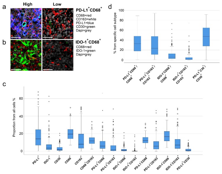
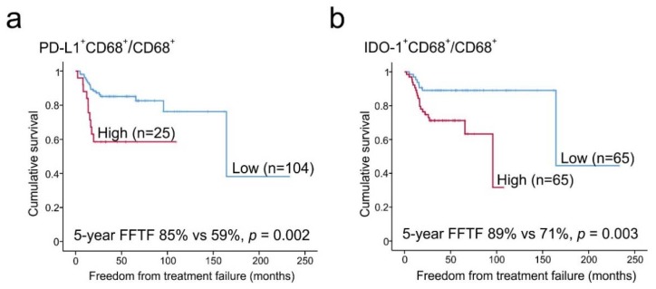
Tumor microenvironment and immune escape affect pathogenesis and survival in classical Hodgkin lymphoma (cHL). While tumor-associated macrophage (TAM) content has been associated with poor outcomes, macrophage-derived determinants with clinical impact have remained undefined. Here, we have used multiplex immunohistochemistry and digital image analysis to characterize TAM immunophenotypes with regard to expression of checkpoint molecules programmed cell death ligand 1 (PD-L1) and indoleamine 2,3-dioxygenase 1 (IDO-1) from the diagnostic tumor tissue samples of 130 cHL patients, and correlated the findings with clinical characteristics and survival. We show that a large proportion of TAMs express PD-L1 (CD68, median 32%; M2 type CD163, median 22%), whereas the proportion of TAMs expressing IDO-1 is lower (CD68, median 5.5%; CD163, median 1.4%). A high proportion of PD-L1 and IDO-1 expressing TAMs from all TAMs (CD68), or from CD163 TAMs, is associated with inferior outcome. In multivariate analysis with age and stage, high proportions of PD-L1 and IDO-1 TAMs remain independent prognostic factors for freedom from treatment failure (PD-L1CD68/CD68, HR = 2.63, 95% CI 1.17-5.88, = 0.019; IDO-1CD68/CD68, HR = 2.48, 95% CI 1.03-5.95, = 0.042). In contrast, proportions of PD-L1 tumor cells, all TAMs or PD-L1 and IDO-1 TAMs are not associated with outcome. The findings implicate that adverse prognostic impact of TAMs is checkpoint-dependent in cHL.
肿瘤微环境和免疫逃逸影响经典型霍奇金淋巴瘤(cHL)的发病机制和生存率。虽然肿瘤相关巨噬细胞(TAM)含量与不良预后相关,但具有临床影响的巨噬细胞衍生决定因素仍不明确。在此,我们使用多重免疫组织化学和数字图像分析,从130例cHL患者的诊断性肿瘤组织样本中,对TAM免疫表型进行了关于检查点分子程序性细胞死亡配体1(PD-L1)和吲哚胺2,3-双加氧酶1(IDO-1)表达的特征分析,并将结果与临床特征和生存率相关联。我们发现,很大一部分TAM表达PD-L1(CD68,中位数32%;M2型CD163,中位数22%),而表达IDO-1的TAM比例较低(CD68,中位数5.5%;CD163,中位数1.4%)。所有TAM(CD68)或CD163 TAM中高比例表达PD-L1和IDO-1的TAM与较差的预后相关。在年龄和分期的多变量分析中,高比例的PD-L1和IDO-1 TAM仍然是免于治疗失败的独立预后因素(PD-L1CD68/CD68,HR = 2.63,95% CI 1.17-5.88,P = 0.019;IDO-1CD68/CD68,HR = 2.48,95% CI 1.03-5.95,P = 0.042)。相比之下,PD-L1肿瘤细胞、所有TAM或PD-L1和IDO-1 TAM的比例与预后无关。这些发现表明,在cHL中,TAM的不良预后影响是检查点依赖性的。