Biosciences, College of Health and Life Sciences, Brunel University London, Uxbridge, United Kingdom.
School of Biosciences and Technology, Vellore Institute of Technology, Vellore, India.
Front Immunol. 2020 Mar 25;11:355. doi: 10.3389/fimmu.2020.00355. eCollection 2020.
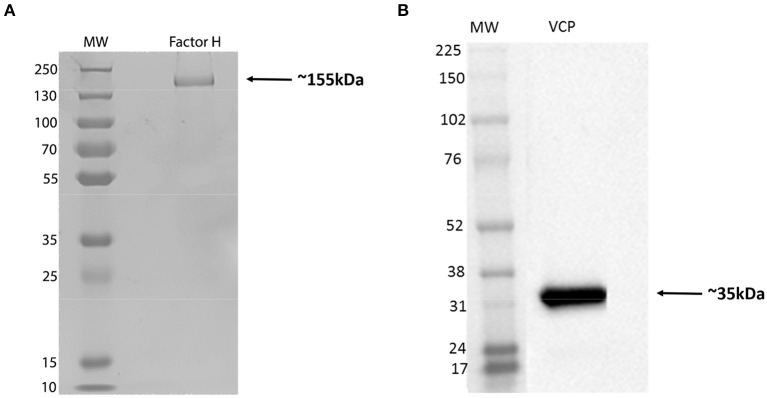
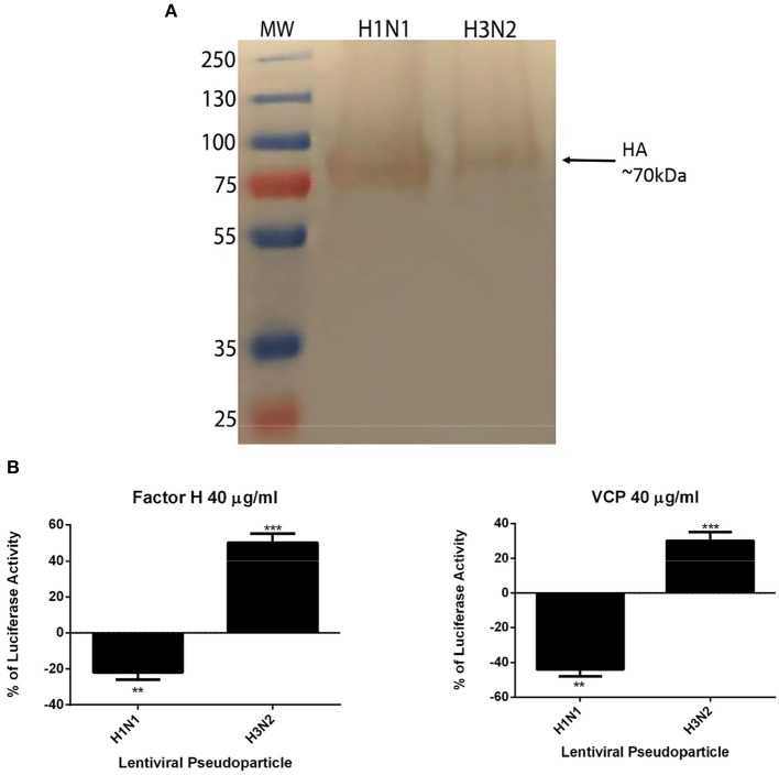
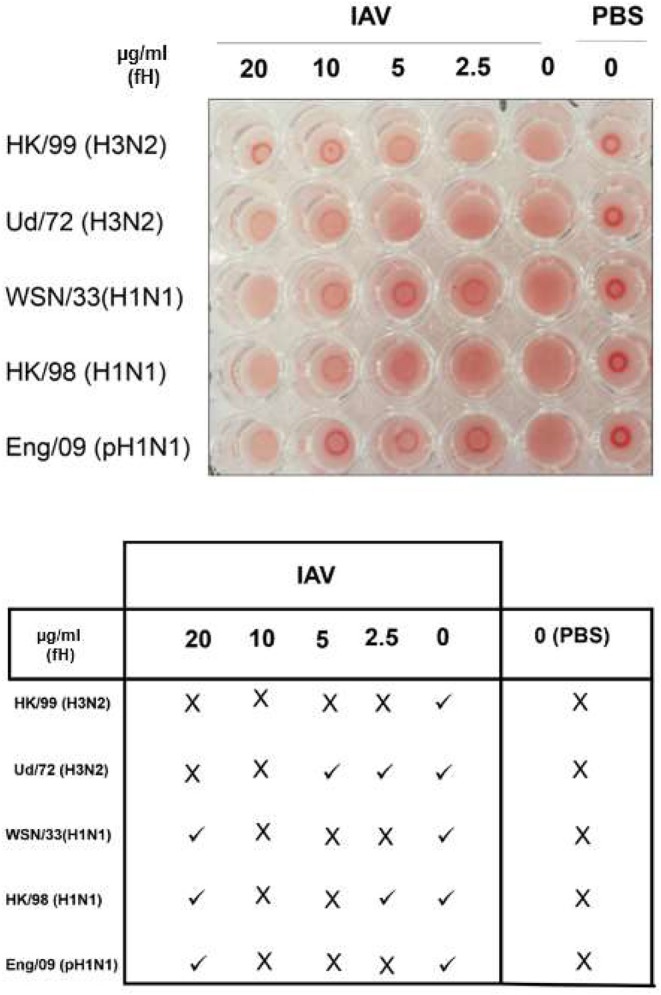
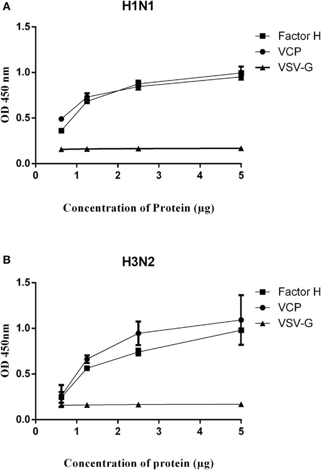
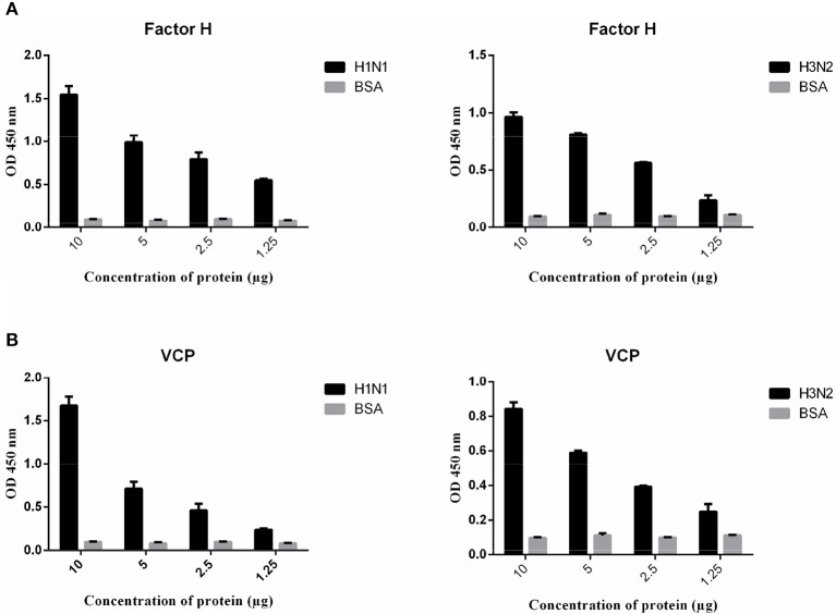
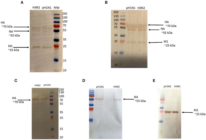
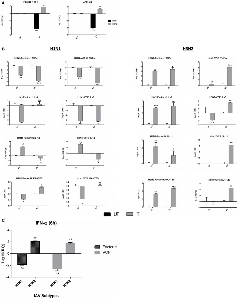
The complement system is an ancient innate immune defense mechanism that can recognize molecular patterns on the invading pathogens. Factor H, as an inhibitor of the alternative pathway, down-regulates complement activation on the host cell surface. Locally synthesized factor H at the site of infection/injury, including lungs, can act as a pattern recognition molecule without involving complement activation. Here, we report that factor H, a sialic acid binder, interacts with influenza A virus (IAV) and modulates IAV entry, as evident from down-regulation of matrix protein 1 (M1) in H1N1 subtype-infected cells and up-regulation of M1 expression in H3N2-infected A549 cells. Far-western blot revealed that factor H binds hemagglutinin (HA, 70 kDa), neuraminidase (NA, ~60 kDa), and M1 (25 kDa). IAV-induced transcriptional levels of IFN-α, TNF-α, IL-12, IL-6, IFN-α, and RANTES were reduced following factor H treatment for the H1N1 subtype at 6 h post-infection. However, for the H3N2 subtype, mRNA levels of these pro-inflammatory cytokines were enhanced. A recombinant form of vaccinia virus complement control protein (VCP), which like factor H, contains CCP modules and has complement-regulatory activity, mirrored the results obtained with factor H. Both factor H (25%), and VCP (45%) were found to reduce luciferase reporter activity in MDCK cells transduced with H1N1 pseudotyped lentiviral particles. Factor H (50%) and VCP (30%) enhanced the luciferase reporter activity for H3N2, suggesting an entry inhibitory role of factor H and VCP against H1N1, but not H3N2. Thus, factor H can modulate IAV infection and inflammatory responses, independent of its complement-related functions.
补体系统是一种古老的先天免疫防御机制,能够识别入侵病原体上的分子模式。因子 H 作为旁路途径的抑制剂,下调宿主细胞表面的补体激活。在感染/损伤部位合成的局部因子 H,包括肺部,可以作为一种模式识别分子,而不涉及补体激活。在这里,我们报告因子 H(一种唾液酸结合蛋白)与甲型流感病毒(IAV)相互作用并调节 IAV 进入,这从 H1N1 亚型感染细胞中基质蛋白 1(M1)的下调和 H3N2 感染的 A549 细胞中 M1 表达的上调中可以明显看出。远 Western blot 显示因子 H 结合血凝素(HA,70 kDa)、神经氨酸酶(NA,60 kDa)和 M1(~25 kDa)。在 H1N1 亚型感染后 6 小时,因子 H 处理可降低 IAV 诱导的 IFN-α、TNF-α、IL-12、IL-6、IFN-α 和 RANTES 的转录水平。然而,对于 H3N2 亚型,这些促炎细胞因子的 mRNA 水平增强。含有 CCP 模块并具有补体调节活性的痘苗病毒补体控制蛋白(VCP)的重组形式与因子 H 的结果相似。在转导 H1N1 假型慢病毒颗粒的 MDCK 细胞中,因子 H(25%)和 VCP(45%)均降低荧光素酶报告基因活性。因子 H(50%)和 VCP(30%)增强了 H3N2 的荧光素酶报告基因活性,表明因子 H 和 VCP 对 H1N1 具有抑制作用,但对 H3N2 没有抑制作用。因此,因子 H 可以调节 IAV 感染和炎症反应,而不依赖其补体相关功能。