Department of Women's and Children's Health, Karolinska Institutet, Stockholm, Sweden.
Department of Obstetrics and Gynecology, Shiga University of Medical Science, Seta Tsukinowa-cho, Otsu City, Shiga, 520-2192, Japan.
J Neuroinflammation. 2020 Apr 11;17(1):111. doi: 10.1186/s12974-020-01792-7.
Neuroinflammation plays an important role in neonatal hypoxic-ischemic encephalopathy (HIE). Although microglia are largely responsible for injury-induced inflammatory response, they play beneficial roles in both normal and disease states. However, the effects of microglial depletion on neonatal HIE remain unclear.
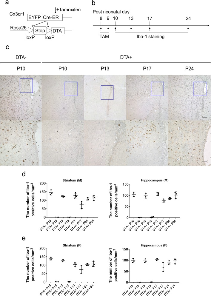
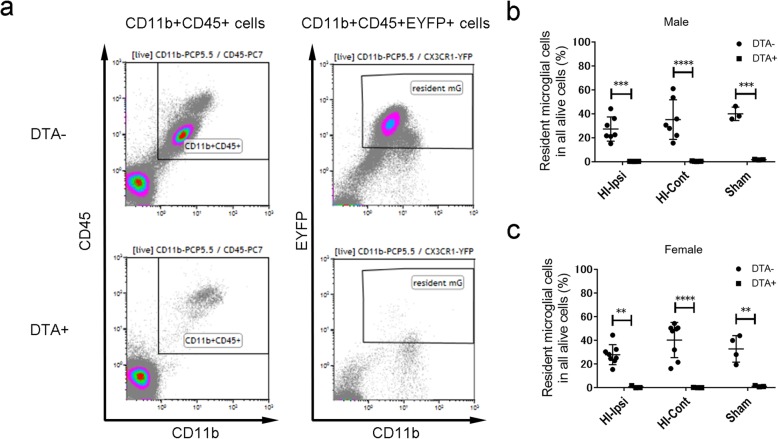
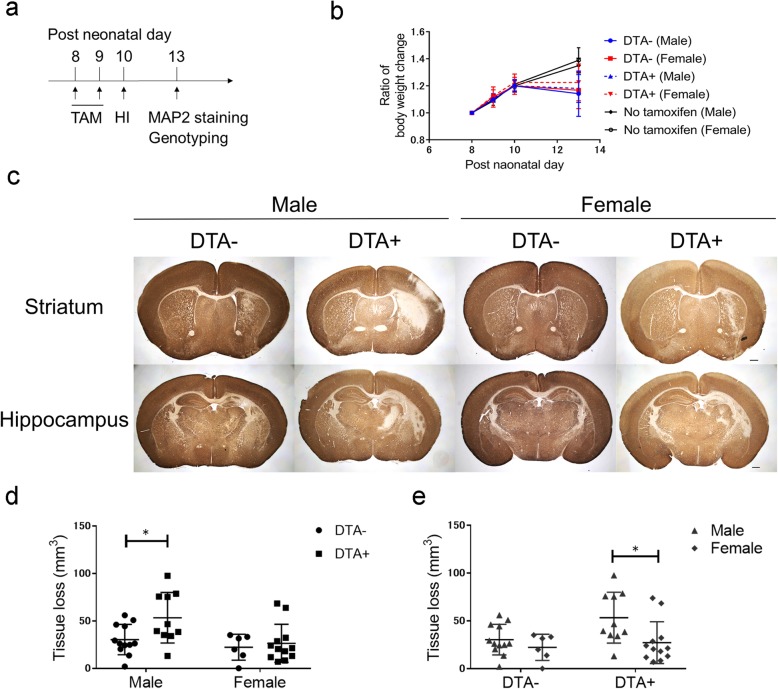
Tamoxifen was administered to Cx3cr1Rosa26 (microglia-depleted model) and Cx3cr1Rosa26 (control) mice at P8 and P9 to assess the effect of microglial depletion. The density of microglia was quantified using Iba-1 staining. Moreover, the proportion of resident microglia after the HI insult was analyzed using flow cytometric analysis. At P10, the HI insult was conducted using the Rice-Vannucci procedure at P10. The infarct size and apoptotic cells were analyzed at P13. Cytokine analyses were performed using quantitative polymerase chain reaction and enzyme-linked immunosorbent assay (ELISA) at P13.
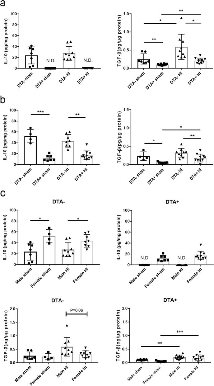
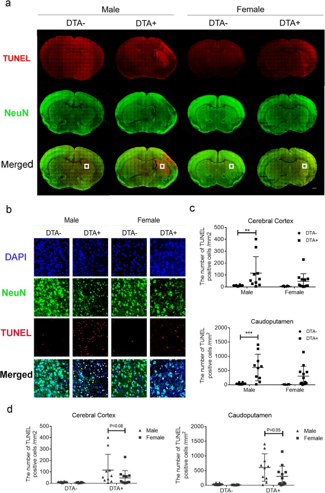
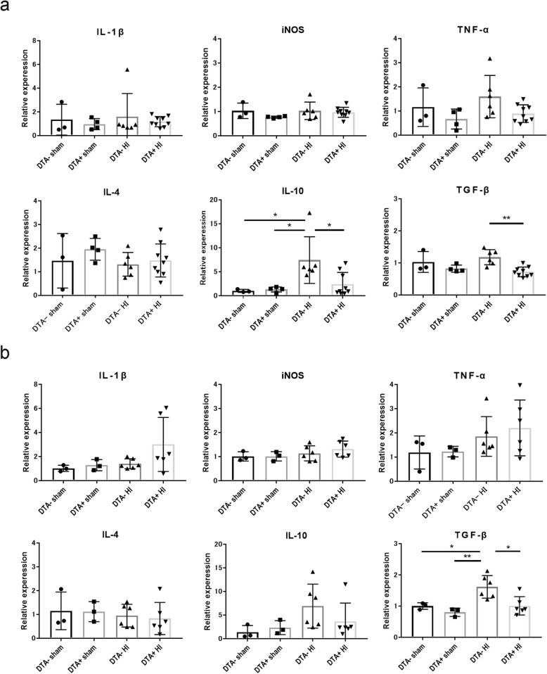
At P10, tamoxifen administration induced > 99% microglial depletion in DTA mice. Following HI insult, there was persisted microglial depletion over 97% at P13. Compared to male DTA mice, male DTA mice exhibited significantly larger infarct volumes; however, there were no significant differences among females. Moreover, compared to male DTA mice, male DTA mice had a significantly higher density of TUNEL cells in the caudoputamen, cerebral cortex, and thalamus. Moreover, compared to female DTA mice, female DTA mice showed a significantly greater number of TUNEL cells in the hippocampus and thalamus. Compared to DTA mice, ELISA revealed significantly lower IL-10 and TGF-β levels in both male and female DTA mice under both normal conditions and after HI (more pronounced).
We established a microglial depletion model that aggravated neuronal damage and apoptosis after the HI insult, which was predominantly observed in males.
神经炎症在新生儿缺氧缺血性脑病(HIE)中起着重要作用。虽然小胶质细胞在很大程度上负责损伤诱导的炎症反应,但它们在正常和疾病状态下都发挥着有益的作用。然而,小胶质细胞耗竭对新生儿 HIE 的影响尚不清楚。
在 P8 和 P9 时用他莫昔芬处理 Cx3cr1Rosa26(小胶质细胞耗竭模型)和 Cx3cr1Rosa26(对照)小鼠,以评估小胶质细胞耗竭的效果。用 Iba-1 染色来量化小胶质细胞的密度。此外,用流式细胞术分析 HI 损伤后驻留小胶质细胞的比例。在 P10 时,用 Rice-Vannucci 法进行 HI 损伤。在 P13 时分析梗死面积和凋亡细胞。在 P13 时用定量聚合酶链反应和酶联免疫吸附试验(ELISA)进行细胞因子分析。
在 P10 时,他莫昔芬给药诱导 DTA 小鼠的小胶质细胞耗竭超过 99%。在 HI 损伤后,P13 时仍有超过 97%的小胶质细胞耗竭。与雄性 DTA 小鼠相比,雄性 DTA 小鼠的梗死体积明显更大;然而,雌性之间没有显著差异。此外,与雄性 DTA 小鼠相比,雄性 DTA 小鼠的尾壳核、大脑皮层和丘脑的 TUNEL 细胞密度明显更高。此外,与雌性 DTA 小鼠相比,雌性 DTA 小鼠的海马体和丘脑的 TUNEL 细胞数量明显更多。与 DTA 小鼠相比,ELISA 显示在正常情况下和 HI 后(更明显),雄性和雌性 DTA 小鼠的 IL-10 和 TGF-β水平均明显降低。
我们建立了一个小胶质细胞耗竭模型,该模型在 HI 损伤后加重了神经元损伤和凋亡,主要发生在雄性中。