• 文献检索
  • 文档翻译
  • 深度研究
  • 学术资讯
  • Suppr Zotero 插件Zotero 插件
  • 邀请有礼
  • 套餐&价格
  • 历史记录
应用&插件
Suppr Zotero 插件Zotero 插件浏览器插件Mac 客户端Windows 客户端微信小程序
定价
高级版会员购买积分包购买API积分包
服务
文献检索文档翻译深度研究API 文档MCP 服务
关于我们
关于 Suppr公司介绍联系我们用户协议隐私条款
关注我们

Suppr 超能文献

核心技术专利:CN118964589B侵权必究
粤ICP备2023148730 号-1Suppr @ 2026

文献检索

告别复杂PubMed语法,用中文像聊天一样搜索,搜遍4000万医学文献。AI智能推荐,让科研检索更轻松。

立即免费搜索

文件翻译

保留排版,准确专业,支持PDF/Word/PPT等文件格式,支持 12+语言互译。

免费翻译文档

深度研究

AI帮你快速写综述,25分钟生成高质量综述,智能提取关键信息,辅助科研写作。

立即免费体验

局部晚期宫颈癌的 DCE-MRI:Tofts 分析与非模型分析的比较。

DCE-MRI of locally-advanced carcinoma of the uterine cervix: Tofts analysis versus non-model-based analyses.

机构信息

Department of Radiation Biology, Institute for Cancer Research, Oslo University Hospital, Oslo, Norway.

Department of Radiology and Nuclear Medicine, Oslo University Hospital, Oslo, Norway.

出版信息

Radiat Oncol. 2020 Apr 15;15(1):79. doi: 10.1186/s13014-020-01526-2.

DOI:10.1186/s13014-020-01526-2
PMID:32293487
原文链接:https://pmc.ncbi.nlm.nih.gov/articles/PMC7158049/
Abstract

BACKGROUND

Dynamic contrast-enhanced magnetic resonance imaging (DCE-MRI) may provide biomarkers of the outcome of locally-advanced cervical carcinoma (LACC). There is, however, no agreement on how DCE-MR recordings should be analyzed. Previously, we have analyzed DCE-MRI data of LACC using non-model-based strategies. In the current study, we analyzed DCE-MRI data of LACC using the Tofts pharmacokinetic model, and the biomarkers derived from this analysis were compared with those derived from the non-model-based analyses.

METHODS

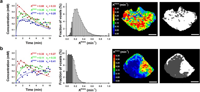
Eighty LACC patients given cisplatin-based chemoradiotherapy with curative intent were included in the study. Treatment outcome was recorded as disease-free survival (DFS) and overall survival (OS). DCE-MRI series were analyzed voxelwise to produce K and v frequency distributions, and ROC analysis was used to identify the parameters of the frequency distributions having the greatest potential as biomarkers. The prognostic power of these parameters was compared with that of the non-model-based parameters LETV (low-enhancing tumor volume) and TVIS (tumor volume with increasing signal).

RESULTS

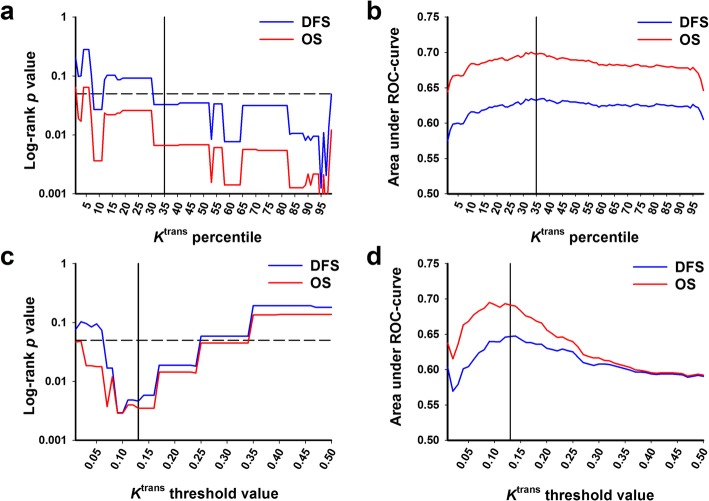
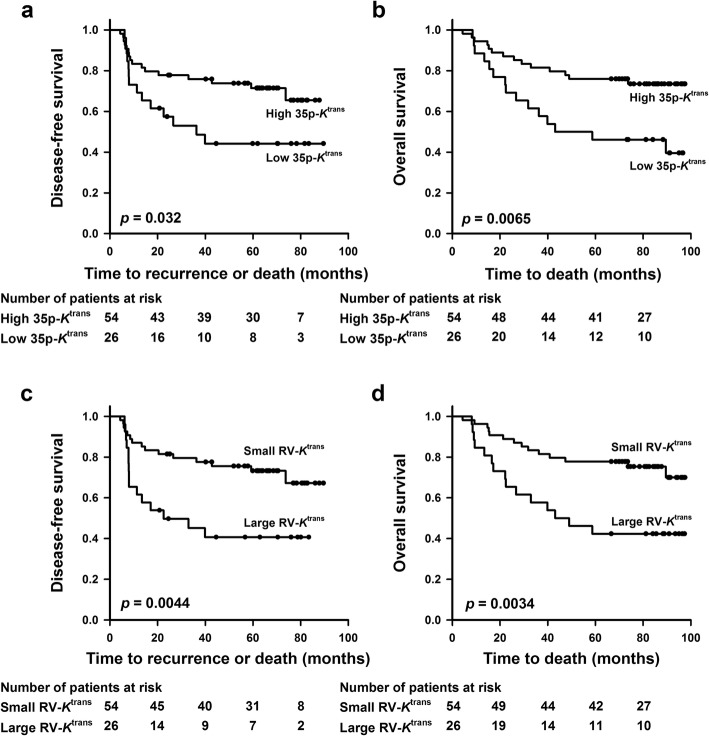
Poor DFS and OS were associated with low values of K, whereas there was no association between treatment outcome and v. The K parameters having the greatest prognostic value were p35-K (the K value at the 35 percentile of a frequency distribution) and RV-K (the tumor subvolume with K values below 0.13 min). Multivariate analysis including clinical parameters and p35-K or RV-K revealed that RV-K was the only independent prognostic factor of DFS and OS. There were significant correlations between RV-K and LETV and between RV-K and TVIS, and the prognostic power of RV-K was similar to that of LETV and TVIS.

CONCLUSIONS

Biomarkers of the outcome of LACC can be provided by analyzing DCE-MRI series using the Tofts pharmacokinetic model. However, these biomarkers do not appear to have greater prognostic value than biomarkers determined by non-model-based analyses.

摘要

背景

动态对比增强磁共振成像(DCE-MRI)可能为局部晚期宫颈癌(LACC)的治疗结果提供生物标志物。然而,目前对于 DCE-MR 记录的分析方法尚未达成共识。先前,我们使用基于非模型的策略分析了 LACC 的 DCE-MRI 数据。在本研究中,我们使用 Tofts 药代动力学模型分析了 LACC 的 DCE-MRI 数据,并将从这种分析中得出的生物标志物与从非模型分析中得出的生物标志物进行了比较。

方法

本研究纳入了 80 例接受顺铂为基础的放化疗治疗的 LACC 患者。治疗结果记录为无病生存率(DFS)和总生存率(OS)。对 DCE-MRI 系列进行体素分析,以生成 K 和 v 频率分布,并使用 ROC 分析来确定作为生物标志物具有最大潜力的频率分布参数。将这些参数的预后能力与非模型参数 LETV(低增强肿瘤体积)和 TVIS(信号增强肿瘤体积)进行比较。

结果

DFS 和 OS 较差与 K 值较低相关,而治疗结果与 v 之间没有相关性。具有最大预后价值的 K 参数是 p35-K(频率分布的第 35 百分位数的 K 值)和 RV-K(K 值低于 0.13 min 的肿瘤亚体积)。包括临床参数和 p35-K 或 RV-K 的多变量分析表明,RV-K 是 DFS 和 OS 的唯一独立预后因素。RV-K 与 LETV 之间以及 RV-K 与 TVIS 之间存在显著相关性,且 RV-K 的预后能力与 LETV 和 TVIS 相似。

结论

使用 Tofts 药代动力学模型分析 DCE-MRI 系列可以提供 LACC 治疗结果的生物标志物。然而,这些生物标志物似乎没有比基于非模型的分析确定的生物标志物具有更大的预后价值。

https://cdn.ncbi.nlm.nih.gov/pmc/blobs/af01/7158049/ce2770d1aad7/13014_2020_1526_Fig5_HTML.jpg
https://cdn.ncbi.nlm.nih.gov/pmc/blobs/af01/7158049/9cb48a433cd9/13014_2020_1526_Fig1_HTML.jpg
https://cdn.ncbi.nlm.nih.gov/pmc/blobs/af01/7158049/1a4b9635de80/13014_2020_1526_Fig2_HTML.jpg
https://cdn.ncbi.nlm.nih.gov/pmc/blobs/af01/7158049/24ed5117907e/13014_2020_1526_Fig3_HTML.jpg
https://cdn.ncbi.nlm.nih.gov/pmc/blobs/af01/7158049/b44392a0f3cb/13014_2020_1526_Fig4_HTML.jpg
https://cdn.ncbi.nlm.nih.gov/pmc/blobs/af01/7158049/ce2770d1aad7/13014_2020_1526_Fig5_HTML.jpg

相似文献

1
DCE-MRI of locally-advanced carcinoma of the uterine cervix: Tofts analysis versus non-model-based analyses.局部晚期宫颈癌的 DCE-MRI:Tofts 分析与非模型分析的比较。
Radiat Oncol. 2020 Apr 15;15(1):79. doi: 10.1186/s13014-020-01526-2.
2
Pharmacokinetic analysis of DCE-MRI data of locally advanced cervical carcinoma with the Brix model.基于 Brix 模型的局部晚期宫颈癌 DCE-MRI 药代动力学分析。
Acta Oncol. 2019 Jun;58(6):828-837. doi: 10.1080/0284186X.2019.1580386. Epub 2019 Feb 27.
3
Pharmacokinetic parameters derived from dynamic contrast enhanced MRI of cervical cancers predict chemoradiotherapy outcome.从宫颈癌动态对比增强 MRI 得出的药代动力学参数可预测放化疗结果。
Radiother Oncol. 2013 Apr;107(1):117-22. doi: 10.1016/j.radonc.2012.11.007. Epub 2013 Jan 17.
4
Voxelwise comparison of perfusion parameters estimated using dynamic contrast enhanced (DCE) computed tomography and DCE-magnetic resonance imaging in locally advanced cervical cancer.基于动态对比增强 CT 和 DCE 磁共振成像的局部晚期宫颈癌灌注参数的体素比较。
Acta Oncol. 2013 Oct;52(7):1360-8. doi: 10.3109/0284186X.2013.813637. Epub 2013 Sep 5.
5
The prognostic value of dynamic contrast-enhanced MRI contrast agent transfer constant K in cervical cancer is explained by plasma flow rather than vessel permeability.动态对比增强磁共振成像造影剂转移常数K在宫颈癌中的预后价值是由血浆流动而非血管通透性所解释的。
Br J Cancer. 2017 May 23;116(11):1436-1443. doi: 10.1038/bjc.2017.121. Epub 2017 Apr 27.
6
DCE-MRI-Derived Measures of Tumor Hypoxia and Interstitial Fluid Pressure Predict Outcomes in Cervical Carcinoma.DCE-MRI 衍生的肿瘤乏氧和间质液压力测量预测宫颈癌结局。
Int J Radiat Oncol Biol Phys. 2018 Nov 15;102(4):1193-1201. doi: 10.1016/j.ijrobp.2018.04.035. Epub 2018 Apr 19.
7
Pretreatment late-phase DCE-MRI predicts outcome in locally advanced cervix cancer.治疗前晚期动态对比增强磁共振成像可预测局部晚期宫颈癌的预后。
Acta Oncol. 2017 May;56(5):675-681. doi: 10.1080/0284186X.2017.1294762. Epub 2017 Feb 28.
8
Short-term pretreatment DCE-MRI in prediction of outcome in locally advanced cervical cancer.短期预处理动态对比增强磁共振成像在预测局部晚期宫颈癌预后中的应用
Radiother Oncol. 2015 Jun;115(3):379-85. doi: 10.1016/j.radonc.2015.05.001. Epub 2015 May 18.
9
Added-value of dynamic contrast-enhanced MRI on prediction of tumor recurrence in locally advanced cervical cancer treated with chemoradiotherapy.动态对比增强磁共振成像在预测接受放化疗的局部晚期宫颈癌肿瘤复发中的附加价值。
Eur Radiol. 2022 Apr;32(4):2529-2539. doi: 10.1007/s00330-021-08279-w. Epub 2021 Oct 13.
10
Preclinical evaluation of Gd-DTPA and gadomelitol as contrast agents in DCE-MRI of cervical carcinoma interstitial fluid pressure.临床前评估 Gd-DTPA 和 gadomelitol 作为宫颈癌间质液压力 DCE-MRI 的对比剂。
BMC Cancer. 2012 Nov 22;12:544. doi: 10.1186/1471-2407-12-544.

引用本文的文献

1
Correlation study of functional magnetic resonance index and clinicopathological features of rectal cancer.直肠癌功能磁共振指数与临床病理特征的相关性研究。
Abdom Radiol (NY). 2024 Jul;49(7):2368-2386. doi: 10.1007/s00261-024-04375-9. Epub 2024 Jun 13.
2
The Utility of Contrast-Enhanced Magnetic Resonance Imaging in Uterine Cervical Cancer: A Systematic Review.对比增强磁共振成像在子宫颈癌中的应用:一项系统评价
Life (Basel). 2023 Jun 12;13(6):1368. doi: 10.3390/life13061368.
3
Dynamic contrast-enhanced MRI in malignant pleural mesothelioma: prediction of outcome based on DCE-MRI measurements in patients undergoing cytotoxic chemotherapy.

本文引用的文献

1
Positron emission tomography with computed tomography imaging (PET/CT) for the radiotherapy planning definition of the biological target volume: PART 2.正电子发射断层扫描与计算机断层成像(PET/CT)在放射治疗生物学靶区定义中的应用:第 2 部分。
Crit Rev Oncol Hematol. 2019 Jul;139:117-124. doi: 10.1016/j.critrevonc.2019.03.008. Epub 2019 Mar 20.
2
DCE-MRI and Quantitative Histology Reveal Enhanced Vessel Maturation but Impaired Perfusion and Increased Hypoxia in Bevacizumab-Treated Cervical Carcinoma.DCE-MRI 和定量组织学揭示贝伐珠单抗治疗宫颈癌中增强的血管成熟度,但灌注受损和缺氧增加。
Int J Radiat Oncol Biol Phys. 2019 Jul 1;104(3):666-676. doi: 10.1016/j.ijrobp.2019.03.002. Epub 2019 Mar 8.
3
动态对比增强 MRI 对恶性胸膜间皮瘤的评估:细胞毒性化疗患者的 DCE-MRI 测量值对预后的预测价值
BMC Cancer. 2022 Feb 20;22(1):191. doi: 10.1186/s12885-022-09277-x.
4
Role of MRI-Based Functional Imaging in Improving the Therapeutic Index of Radiotherapy in Cancer Treatment.基于磁共振成像的功能成像在提高癌症放疗治疗指数中的作用。
Front Oncol. 2021 Aug 27;11:645177. doi: 10.3389/fonc.2021.645177. eCollection 2021.
5
Assessment of Hypoxic Tissue Fraction and Prediction of Survival in Cervical Carcinoma by Dynamic Contrast-Enhanced MRI.动态对比增强磁共振成像评估宫颈癌缺氧组织分数及预测生存情况
Front Oncol. 2021 May 20;11:668916. doi: 10.3389/fonc.2021.668916. eCollection 2021.
6
DCE-MRI of Tumor Hypoxia and Hypoxia-Associated Aggressiveness.肿瘤缺氧及缺氧相关侵袭性的动态对比增强磁共振成像
Cancers (Basel). 2020 Jul 20;12(7):1979. doi: 10.3390/cancers12071979.
Pharmacokinetic analysis of DCE-MRI data of locally advanced cervical carcinoma with the Brix model.
基于 Brix 模型的局部晚期宫颈癌 DCE-MRI 药代动力学分析。
Acta Oncol. 2019 Jun;58(6):828-837. doi: 10.1080/0284186X.2019.1580386. Epub 2019 Feb 27.
4
Positron emission tomography with computed tomography imaging (PET/CT) for the radiotherapy planning definition of the biological target volume: PART 1.正电子发射断层扫描与计算机断层成像(PET/CT)在放射治疗计划生物靶区定义中的应用:第 1 部分。
Crit Rev Oncol Hematol. 2019 Aug;140:74-79. doi: 10.1016/j.critrevonc.2019.01.011. Epub 2019 Jan 25.
5
DCE-MRI of patient-derived xenograft models of uterine cervix carcinoma: associations with parameters of the tumor microenvironment.宫颈癌患者来源异种移植模型的 DCE-MRI:与肿瘤微环境参数的相关性。
J Transl Med. 2017 Nov 3;15(1):225. doi: 10.1186/s12967-017-1331-4.
6
Hypoxia in cervical cancer: from biology to imaging.宫颈癌中的缺氧:从生物学到影像学
Clin Transl Imaging. 2017;5(4):373-388. doi: 10.1007/s40336-017-0238-7. Epub 2017 Jul 10.
7
The prognostic value of dynamic contrast-enhanced MRI contrast agent transfer constant K in cervical cancer is explained by plasma flow rather than vessel permeability.动态对比增强磁共振成像造影剂转移常数K在宫颈癌中的预后价值是由血浆流动而非血管通透性所解释的。
Br J Cancer. 2017 May 23;116(11):1436-1443. doi: 10.1038/bjc.2017.121. Epub 2017 Apr 27.
8
Pretreatment late-phase DCE-MRI predicts outcome in locally advanced cervix cancer.治疗前晚期动态对比增强磁共振成像可预测局部晚期宫颈癌的预后。
Acta Oncol. 2017 May;56(5):675-681. doi: 10.1080/0284186X.2017.1294762. Epub 2017 Feb 28.
9
Short-term pretreatment DCE-MRI in prediction of outcome in locally advanced cervical cancer.短期预处理动态对比增强磁共振成像在预测局部晚期宫颈癌预后中的应用
Radiother Oncol. 2015 Jun;115(3):379-85. doi: 10.1016/j.radonc.2015.05.001. Epub 2015 May 18.
10
Relationship between intravoxel incoherent motion diffusion-weighted MRI and dynamic contrast-enhanced MRI in tissue perfusion of cervical cancers.体素内不相干运动扩散加权磁共振成像与动态对比增强磁共振成像在宫颈癌组织灌注中的关系
J Magn Reson Imaging. 2015 Aug;42(2):454-9. doi: 10.1002/jmri.24808. Epub 2014 Nov 21.