Department of Obstetrics and Gynecology, Qilu Hospital of Shandong University, Jinan, 250012, Shandong, People's Republic of China.
Department of Cardiology, Qilu Hospital of Shandong University, Jinan, 250012, Shandong, People's Republic of China.
Stem Cell Res Ther. 2020 Mar 23;11(1):133. doi: 10.1186/s13287-020-01639-1.
In our previous research, we found that mesenchymal stem cell (MSC) transplantation therapy can inhibit intimal hyperplasia and enhance endothelial function in arterialized vein grafts in rats. However, whether MSC-derived exosomes (MSC-exosomes) can reduce neointimal formation and its possible mechanism is still unclear.
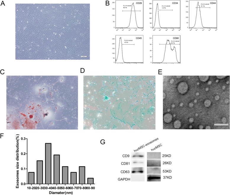
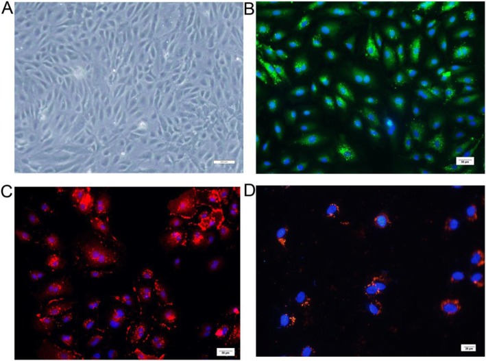
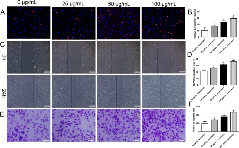
The primary human umbilical cord MSCs (hucMSCs) and human umbilical vein endothelial cells (HUVECs) were isolated and characterized by flow cytometry and immunofluorescence. The exosomes derived from hucMSCs (hucMSC-exosomes) were identified by transmission electron microscopy and western blots. hucMSC-exosomes were intravenously injected into a rat model of vein grafting, and its effect on vein grafts reendothelialization and intimal hyperplasia was assessed by physical, histological, immunohistochemistry, and immunofluorescence examinations. The effects of hucMSC-exosomes on endothelial cells were evaluated by integrated experiment, EdU staining, scratch assay, and Transwell assay. The expression levels of key gene and pathways associated with the biological activity of vascular endothelial cells were evaluated following the stimulation of hucMSC-exosomes.
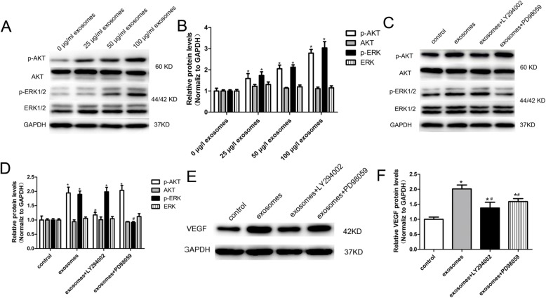
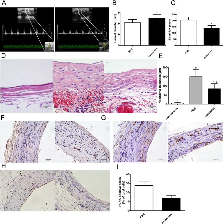
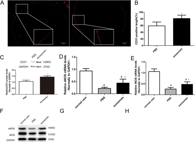
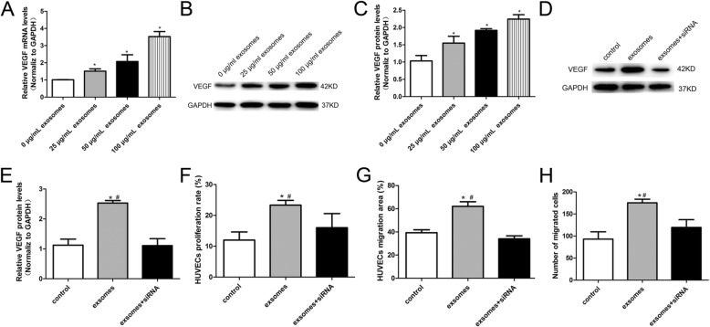
We successfully isolated and characterized primary hucMSCs and hucMSC-exosomes and primary HUVECs. We verified that the systemic administration of hucMSC-exosomes accelerates reendothelialization and decreases intimal hyperplasia of autologous vein graft in a rat model. We also identified that hucMSC-exosomes can be uptaken by endothelial cells to stimulate cell proliferative and migratory activity in vitro. Furthermore, we detected that vascular endothelial growth factor (VEGF) plays an important part in hucMSC-exosome-mediated proliferation and migration in HUVECs. In addition, we also provided evidence that the signalling pathways of PI3K/AKT and MAPK/ERK1/2 take part in hucMSC-exosome-induced VEGF regulation.
Our data suggest that hucMSC-exosomes exert a vasculoprotective role in the setting of vein graft disease, which may provide a new clue to protect against vein graft failure in the future.
在我们之前的研究中,我们发现间充质干细胞(MSC)移植治疗可以抑制大鼠动脉化静脉移植物的内膜增生并增强内皮功能。然而,MSC 衍生的外泌体(MSC-exosomes)是否可以减少新生内膜形成及其可能的机制尚不清楚。
原代人脐带 MSC(hucMSCs)和人脐静脉内皮细胞(HUVECs)通过流式细胞术和免疫荧光法分离和鉴定。通过透射电子显微镜和 Western blot 鉴定 hucMSCs 衍生的外泌体(hucMSC-exosomes)。将 hucMSC-exosomes 静脉内注射到大鼠静脉移植模型中,通过物理、组织学、免疫组织化学和免疫荧光检查评估其对静脉移植物再内皮化和内膜增生的影响。通过整合实验、EdU 染色、划痕实验和 Transwell 实验评估 hucMSC-exosomes 对内皮细胞的影响。评估 hucMSC-exosomes 刺激后与血管内皮细胞生物学活性相关的关键基因和途径的表达水平。
我们成功分离和鉴定了原代 hucMSCs 和 hucMSC-exosomes 以及原代 HUVECs。我们验证了全身给予 hucMSC-exosomes 可加速大鼠自体静脉移植物的再内皮化并减少内膜增生。我们还发现 hucMSC-exosomes 可被内皮细胞摄取,以刺激体外细胞增殖和迁移活性。此外,我们检测到血管内皮生长因子(VEGF)在 hucMSC-exosome 介导的 HUVEC 增殖和迁移中起重要作用。此外,我们还提供了证据表明,PI3K/AKT 和 MAPK/ERK1/2 信号通路参与了 hucMSC-exosome 诱导的 VEGF 调节。
我们的数据表明,hucMSC-exosomes 在静脉移植物疾病中发挥血管保护作用,这可能为未来防止静脉移植物失败提供新的线索。