• 文献检索
  • 文档翻译
  • 深度研究
  • 学术资讯
  • Suppr Zotero 插件Zotero 插件
  • 邀请有礼
  • 套餐&价格
  • 历史记录
应用&插件
Suppr Zotero 插件Zotero 插件浏览器插件Mac 客户端Windows 客户端微信小程序
定价
高级版会员购买积分包购买API积分包
服务
文献检索文档翻译深度研究API 文档MCP 服务
关于我们
关于 Suppr公司介绍联系我们用户协议隐私条款
关注我们

Suppr 超能文献

核心技术专利:CN118964589B侵权必究
粤ICP备2023148730 号-1Suppr @ 2026

文献检索

告别复杂PubMed语法,用中文像聊天一样搜索,搜遍4000万医学文献。AI智能推荐,让科研检索更轻松。

立即免费搜索

文件翻译

保留排版,准确专业,支持PDF/Word/PPT等文件格式,支持 12+语言互译。

免费翻译文档

深度研究

AI帮你快速写综述,25分钟生成高质量综述,智能提取关键信息,辅助科研写作。

立即免费体验

基于方向局部对比度的机器学习在彩色眼底图像中微动脉瘤的检测。

Microaneurysms detection in color fundus images using machine learning based on directional local contrast.

机构信息

College of Computer Science and Technology, Zhejiang University of Technology, Hangzhou, 310023, China.

Research Center of Intelligent Healthcare, Faculty of Health and Life Science, Coventry University, Coventry, CV1 5RW, UK.

出版信息

Biomed Eng Online. 2020 Apr 15;19(1):21. doi: 10.1186/s12938-020-00766-3.

DOI:10.1186/s12938-020-00766-3
PMID:32295576
原文链接:https://pmc.ncbi.nlm.nih.gov/articles/PMC7161183/
Abstract

BACKGROUND

As one of the major complications of diabetes, diabetic retinopathy (DR) is a leading cause of visual impairment and blindness due to delayed diagnosis and intervention. Microaneurysms appear as the earliest symptom of DR. Accurate and reliable detection of microaneurysms in color fundus images has great importance for DR screening.

METHODS

A microaneurysms' detection method using machine learning based on directional local contrast (DLC) is proposed for the early diagnosis of DR. First, blood vessels were enhanced and segmented using improved enhancement function based on analyzing eigenvalues of Hessian matrix. Next, with blood vessels excluded, microaneurysm candidate regions were obtained using shape characteristics and connected components analysis. After image segmented to patches, the features of each microaneurysm candidate patch were extracted, and each candidate patch was classified into microaneurysm or non-microaneurysm. The main contributions of our study are (1) making use of directional local contrast in microaneurysms' detection for the first time, which does make sense for better microaneurysms' classification. (2) Applying three different machine learning techniques for classification and comparing their performance for microaneurysms' detection. The proposed algorithm was trained and tested on e-ophtha MA database, and further tested on another independent DIARETDB1 database. Results of microaneurysms' detection on the two databases were evaluated on lesion level and compared with existing algorithms.

RESULTS

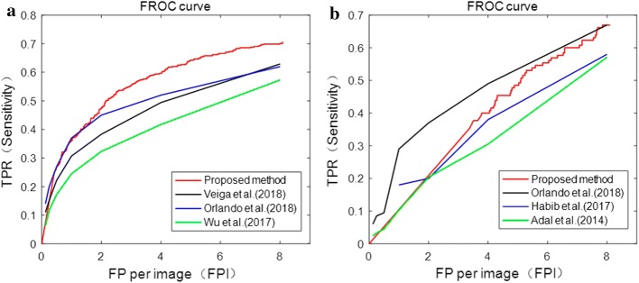
The proposed method has achieved better performance compared with existing algorithms on accuracy and computation time. On e-ophtha MA and DIARETDB1 databases, the area under curve (AUC) of receiver operating characteristic (ROC) curve was 0.87 and 0.86, respectively. The free-response ROC (FROC) score on the two databases was 0.374 and 0.210, respectively. The computation time per image with resolution of 2544×1969, 1400×960 and 1500×1152 is 29 s, 3 s and 2.6 s, respectively.

CONCLUSIONS

The proposed method using machine learning based on directional local contrast of image patches can effectively detect microaneurysms in color fundus images and provide an effective scientific basis for early clinical DR diagnosis.

摘要

背景

糖尿病性视网膜病变(DR)是糖尿病的主要并发症之一,由于诊断和干预不及时,它是导致视力损害和失明的主要原因。微动脉瘤是 DR 的最早症状。准确可靠地检测彩色眼底图像中的微动脉瘤对于 DR 的筛查具有重要意义。

方法

提出了一种基于方向局部对比度(DLC)的基于机器学习的微动脉瘤检测方法,用于 DR 的早期诊断。首先,使用基于分析 Hessian 矩阵特征值的改进增强函数增强和分割血管。接下来,在排除血管后,使用形状特征和连通分量分析获得微动脉瘤候选区域。图像分割成斑块后,提取每个微动脉瘤候选斑块的特征,并将每个候选斑块分类为微动脉瘤或非微动脉瘤。我们研究的主要贡献是:(1)首次在微动脉瘤检测中使用方向局部对比度,这对更好的微动脉瘤分类很有意义。(2)应用三种不同的机器学习技术进行分类,并比较它们在微动脉瘤检测中的性能。所提出的算法在 e-ophtha MA 数据库上进行训练和测试,并在另一个独立的 DIARETDB1 数据库上进行进一步测试。在病变水平上评估两个数据库上微动脉瘤检测的结果,并与现有算法进行比较。

结果

与现有算法相比,该方法在准确性和计算时间上均具有更好的性能。在 e-ophtha MA 和 DIARETDB1 数据库上,接收器工作特征(ROC)曲线的曲线下面积(AUC)分别为 0.87 和 0.86。两个数据库的自由响应 ROC(FROC)得分分别为 0.374 和 0.210。分辨率为 2544×1969、1400×960 和 1500×1152 的图像的计算时间分别为 29 s、3 s 和 2.6 s。

结论

该方法使用基于图像斑块方向局部对比度的机器学习,可以有效检测彩色眼底图像中的微动脉瘤,为临床早期 DR 诊断提供有效科学依据。

https://cdn.ncbi.nlm.nih.gov/pmc/blobs/90f3/7161183/165946499e87/12938_2020_766_Fig13_HTML.jpg
https://cdn.ncbi.nlm.nih.gov/pmc/blobs/90f3/7161183/705566121943/12938_2020_766_Fig1_HTML.jpg
https://cdn.ncbi.nlm.nih.gov/pmc/blobs/90f3/7161183/4d7f83776c08/12938_2020_766_Fig2_HTML.jpg
https://cdn.ncbi.nlm.nih.gov/pmc/blobs/90f3/7161183/5747db77b968/12938_2020_766_Fig3_HTML.jpg
https://cdn.ncbi.nlm.nih.gov/pmc/blobs/90f3/7161183/8d63e3e7e95e/12938_2020_766_Fig4_HTML.jpg
https://cdn.ncbi.nlm.nih.gov/pmc/blobs/90f3/7161183/54046d9c0509/12938_2020_766_Fig5_HTML.jpg
https://cdn.ncbi.nlm.nih.gov/pmc/blobs/90f3/7161183/b0ff362d9fba/12938_2020_766_Fig6_HTML.jpg
https://cdn.ncbi.nlm.nih.gov/pmc/blobs/90f3/7161183/d98a2c42cba6/12938_2020_766_Fig7_HTML.jpg
https://cdn.ncbi.nlm.nih.gov/pmc/blobs/90f3/7161183/91aed291f0c1/12938_2020_766_Fig8_HTML.jpg
https://cdn.ncbi.nlm.nih.gov/pmc/blobs/90f3/7161183/1b6b5ff3bbe0/12938_2020_766_Fig9_HTML.jpg
https://cdn.ncbi.nlm.nih.gov/pmc/blobs/90f3/7161183/a892ed7ba139/12938_2020_766_Fig10_HTML.jpg
https://cdn.ncbi.nlm.nih.gov/pmc/blobs/90f3/7161183/51c6c4f3b6c0/12938_2020_766_Fig11_HTML.jpg
https://cdn.ncbi.nlm.nih.gov/pmc/blobs/90f3/7161183/638adec1b06f/12938_2020_766_Fig12_HTML.jpg
https://cdn.ncbi.nlm.nih.gov/pmc/blobs/90f3/7161183/165946499e87/12938_2020_766_Fig13_HTML.jpg

相似文献

1
Microaneurysms detection in color fundus images using machine learning based on directional local contrast.基于方向局部对比度的机器学习在彩色眼底图像中微动脉瘤的检测。
Biomed Eng Online. 2020 Apr 15;19(1):21. doi: 10.1186/s12938-020-00766-3.
2
Detection of microaneurysms using ant colony algorithm in the early diagnosis of diabetic retinopathy.使用蚁群算法检测微动脉瘤在糖尿病视网膜病变早期诊断中的应用。
Med Hypotheses. 2019 Aug;129:109242. doi: 10.1016/j.mehy.2019.109242. Epub 2019 May 21.
3
Microaneurysm detection in color eye fundus images for diabetic retinopathy screening.用于糖尿病视网膜病变筛查的彩色眼底图像中的微动脉瘤检测
Comput Biol Med. 2020 Nov;126:103995. doi: 10.1016/j.compbiomed.2020.103995. Epub 2020 Sep 18.
4
Resilient back-propagation machine learning-based classification on fundus images for retinal microaneurysm detection.基于稳健反向传播机器学习的眼底图像视网膜微动脉瘤检测分类。
Int Ophthalmol. 2024 Feb 17;44(1):91. doi: 10.1007/s10792-024-02982-5.
5
Mathematical morphology for microaneurysm detection in fundus images.用于眼底图像中微动脉瘤检测的数学形态学
Eur J Ophthalmol. 2020 Sep;30(5):1135-1142. doi: 10.1177/1120672119843021. Epub 2019 Apr 25.
6
Automatic detection of microaneurysms in retinal fundus images.视网膜眼底图像中微动脉瘤的自动检测。
Comput Med Imaging Graph. 2017 Jan;55:106-112. doi: 10.1016/j.compmedimag.2016.08.001. Epub 2016 Aug 4.
7
Automatic microaneurysm detection in fundus image based on local cross-section transformation and multi-feature fusion.基于局部截面变换和多特征融合的眼底图像自动微动脉瘤检测
Comput Methods Programs Biomed. 2020 Nov;196:105687. doi: 10.1016/j.cmpb.2020.105687. Epub 2020 Aug 8.
8
Microaneurysm Detection Using Principal Component Analysis and Machine Learning Methods.基于主成分分析和机器学习方法的微动脉瘤检测。
IEEE Trans Nanobioscience. 2018 Jul;17(3):191-198. doi: 10.1109/TNB.2018.2840084. Epub 2018 May 24.
9
New hierarchical approach for microaneurysms detection with matched filter and machine learning.基于匹配滤波器和机器学习的微动脉瘤检测新分层方法。
Annu Int Conf IEEE Eng Med Biol Soc. 2015 Aug;2015:4322-5. doi: 10.1109/EMBC.2015.7319351.
10
Hybrid multi-kernel SVM algorithm for detection of microaneurysm in color fundus images.用于彩色眼底图像中微动脉瘤检测的混合多核支持向量机算法
Med Biol Eng Comput. 2022 May;60(5):1377-1390. doi: 10.1007/s11517-022-02534-y. Epub 2022 Mar 24.

引用本文的文献

1
Retinal Microvascular Characteristics-Novel Risk Stratification in Cardiovascular Diseases.视网膜微血管特征——心血管疾病中的新型风险分层
Diagnostics (Basel). 2025 Apr 23;15(9):1073. doi: 10.3390/diagnostics15091073.
2
Perfused and Nonperfused Microaneurysms Identified and Characterized by Structural and Angiographic OCT.通过结构和血管造影光学相干断层扫描识别和表征的灌注与非灌注微动脉瘤
ArXiv. 2023 Oct 9:arXiv:2303.13611v2.
3
Discriminative-Region Multi-Label Classification of Ultra-Widefield Fundus Images.超广角眼底图像的判别区域多标签分类

本文引用的文献

1
FILM: finding the location of microaneurysms on the retina.电影:确定视网膜上微动脉瘤的位置。
Biomed Eng Lett. 2019 Nov 2;9(4):497-506. doi: 10.1007/s13534-019-00136-6. eCollection 2019 Nov.
2
Global and regional diabetes prevalence estimates for 2019 and projections for 2030 and 2045: Results from the International Diabetes Federation Diabetes Atlas, 9 edition.2019 年全球及各区域糖尿病患病率估算值及 2030 年和 2045 年预测值:国际糖尿病联盟糖尿病地图集(第 9 版)的结果。
Diabetes Res Clin Pract. 2019 Nov;157:107843. doi: 10.1016/j.diabres.2019.107843. Epub 2019 Sep 10.
3
Quantitative Ultra-Widefield Angiography and Diabetic Retinopathy Severity: An Assessment of Panretinal Leakage Index, Ischemic Index and Microaneurysm Count.
Bioengineering (Basel). 2023 Sep 6;10(9):1048. doi: 10.3390/bioengineering10091048.
4
Detecting red-lesions from retinal fundus images using unique morphological features.利用独特的形态特征检测眼底图像中的红色病灶。
Sci Rep. 2023 Mar 1;13(1):3487. doi: 10.1038/s41598-023-30459-5.
5
Recent developments on computer aided systems for diagnosis of diabetic retinopathy: a review.糖尿病视网膜病变诊断的计算机辅助系统的最新进展:综述
Multimed Tools Appl. 2023;82(10):14471-14525. doi: 10.1007/s11042-022-13841-9. Epub 2022 Sep 24.
6
Automatic detection of microaneurysms in optical coherence tomography images of retina using convolutional neural networks and transfer learning.利用卷积神经网络和迁移学习自动检测视网膜光学相干断层扫描图像中的微动脉瘤。
Sci Rep. 2022 Aug 17;12(1):13975. doi: 10.1038/s41598-022-18206-8.
7
Review on diabetic retinopathy with deep learning methods.基于深度学习方法的糖尿病视网膜病变综述。
J Med Imaging (Bellingham). 2021 Nov;8(6):060901. doi: 10.1117/1.JMI.8.6.060901. Epub 2021 Nov 29.
8
Red-lesion extraction in retinal fundus images by directional intensity changes' analysis.基于方向强度变化分析的眼底图像红色病灶提取。
Sci Rep. 2021 Sep 14;11(1):18223. doi: 10.1038/s41598-021-97649-x.
定量超广角血管造影与糖尿病视网膜病变严重程度:对全视网膜渗漏指数、缺血指数和微动脉瘤计数的评估。
Ophthalmology. 2019 Nov;126(11):1527-1532. doi: 10.1016/j.ophtha.2019.05.034. Epub 2019 Jun 8.
4
Secondary Observer System for Detection of Microaneurysms in Fundus Images Using Texture Descriptors.基于纹理特征的眼底图像微动脉瘤二级检测观察系统。
J Digit Imaging. 2020 Feb;33(1):159-167. doi: 10.1007/s10278-019-00225-z.
5
Microaneurysm detection in fundus images using a two-step convolutional neural network.使用两步卷积神经网络检测眼底图像中的微动脉瘤。
Biomed Eng Online. 2019 May 29;18(1):67. doi: 10.1186/s12938-019-0675-9.
6
Diabetic retinopathy techniques in retinal images: A review.视网膜图像中的糖尿病性视网膜病变技术:综述。
Artif Intell Med. 2019 Jun;97:168-188. doi: 10.1016/j.artmed.2018.10.009. Epub 2018 Nov 16.
7
Algorithms for red lesion detection in Diabetic Retinopathy: A review.糖尿病视网膜病变中红色病灶检测的算法:综述。
Biomed Pharmacother. 2018 Nov;107:681-688. doi: 10.1016/j.biopha.2018.07.175. Epub 2018 Aug 18.
8
Microaneurysm turnover is a predictor of diabetic retinopathy progression.微动脉瘤的出现与消失与糖尿病视网膜病变的进展有关。
Br J Ophthalmol. 2019 Feb;103(2):222-226. doi: 10.1136/bjophthalmol-2018-311887. Epub 2018 Apr 26.
9
Retinal Microaneurysms Detection Using Local Convergence Index Features.基于局部汇聚指数特征的视网膜微动脉瘤检测
IEEE Trans Image Process. 2018 Jul;27(7):3300-3315. doi: 10.1109/TIP.2018.2815345.
10
Microaneurysm detection using fully convolutional neural networks.基于全卷积神经网络的微动脉瘤检测
Comput Methods Programs Biomed. 2018 May;158:185-192. doi: 10.1016/j.cmpb.2018.02.016. Epub 2018 Feb 22.