Li Zhengqiang, Liu Xianwen, Zhang Quanyin, Zhang Jie, Huang Mingyi, Liu Shuguang
Department of Oral and Maxillofacial Surgery, Stomatological Hospital of Southern Medical University, 366 south of Jiangnan Road, Guangzhou, 510280, China.
BMC Oral Health. 2020 Apr 16;20(1):113. doi: 10.1186/s12903-020-01094-7.
Myxofibrosarcoma (MFS) is a soft tissue sarcoma that commonly occurs in late adult life. It is mainly located in the subcutaneous soft tissues of extremities characterized by a high recurrence rate at the original site. MFS of the head and neck is rare, while it occurs in the maxilla and mandible is extremely rare.
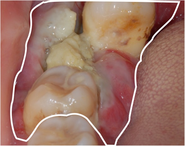
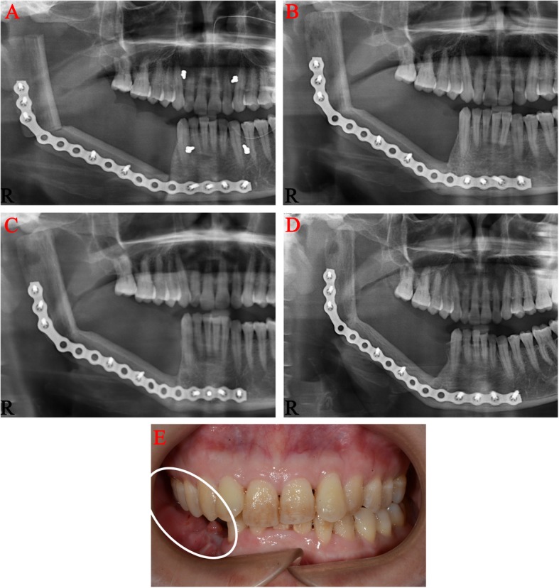
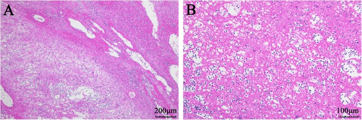
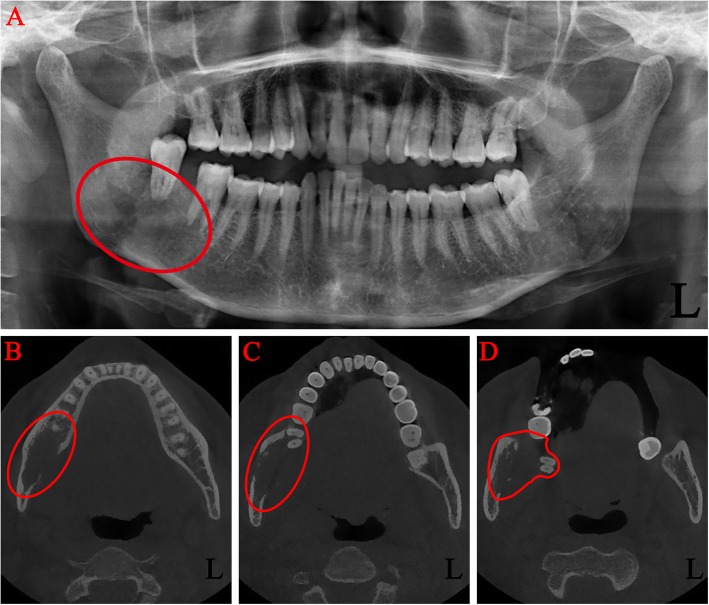
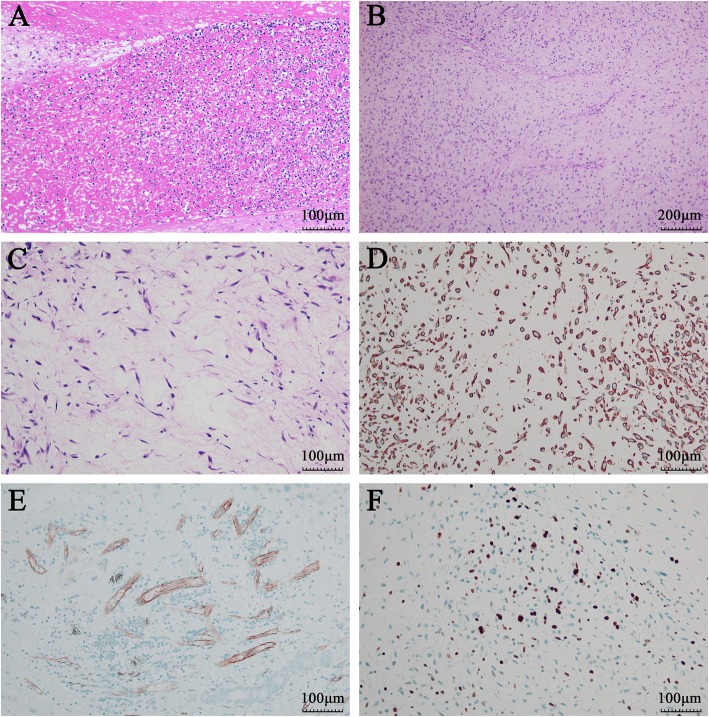
We report a case of MFS of the mandible in a 51-year-old female who presented with a painless gingival swelling and mobile, super-erupted right mandibular second and third molars. Panoramic x-ray and maxillofacial CT revealed an ill-defined radiolucent lesion surrounding the mandibular molars giving a teeth-floating-in-air appearance. Histopathological examination showed scattered spindle and stellate cells with mild atypia distributed in the myxoid stroma. Only a few mitotic figures were identified and no area of tissue necrosis was found. The characteristic thin-walled and curvilinear vasculature were prominent. Immunohistochemistry analysis revealed the tumor cells being positive for vimentin and vascular CD31. CK, S-100, P63, HHF-35 stains were negative. The labeling index of Ki-67 was about 30%. Based on the histopathological and immunohistochemical examinations, the diagnosis of a low-grade MFS was established. This patient underwent a radical segmental excision with a 2-cm margin, supraomohyoid neck dissection and immediate reconstruction of the mandibular continuity defect with a fibular osteocutaneous free flap. This patient has been followed for 20 months to date and has remained disease free.
This report describes a rare case of MFS of the mandible. Recognizing the histopathological features of MFS and applying the appropriate immunohistochemical examinations are crucial in establishing the correct diagnosis. Our case may provide diagnosis and treatment experiences of MFS occurs in the mandible.
黏液纤维肉瘤(MFS)是一种常见于成年人晚期的软组织肉瘤。它主要位于四肢的皮下软组织,其特点是原发部位复发率高。头颈部的MFS较为罕见,而发生在上颌骨和下颌骨的则极为罕见。
我们报告一例51岁女性下颌骨黏液纤维肉瘤病例,患者表现为无痛性牙龈肿胀,右侧下颌第二和第三磨牙松动、伸长。全景X线片和颌面CT显示下颌磨牙周围有边界不清的透射性病变,呈牙齿漂浮在空中的表现。组织病理学检查显示散在的梭形和星状细胞,轻度异型性,分布于黏液样基质中。仅发现少数有丝分裂象,未发现组织坏死区域。特征性的薄壁和曲线状血管明显。免疫组化分析显示肿瘤细胞波形蛋白和血管CD31阳性。细胞角蛋白(CK)、S-100、P63、HHF-35染色均为阴性。Ki-67标记指数约为30%。根据组织病理学和免疫组化检查,确诊为低级别黏液纤维肉瘤。该患者接受了切缘为2cm的根治性节段性切除、肩胛舌骨肌上颈清扫术,并立即用腓骨骨皮瓣修复下颌连续性缺损。该患者至今已随访20个月,无疾病复发。
本报告描述了一例罕见的下颌骨黏液纤维肉瘤病例。认识黏液纤维肉瘤的组织病理学特征并进行适当的免疫组化检查对确立正确诊断至关重要。我们的病例可为下颌骨黏液纤维肉瘤的诊断和治疗提供经验。