Madrakhimov Sanjar Batirovich, Yang Jin Young, Ahn Dong Hyuck, Han Jung Woo, Ha Tae Ho, Park Tae Kwann
Department of Interdisciplinary Program in Biomedical Science, Soonchunhyang Graduate School, Bucheon Hospital, Bucheon, South Korea.
Laboratory for Translational Research on Retinal and Macular Degeneration, Soonchunhyang University Hospital Bucheon, Bucheon, South Korea.
Mol Ther Methods Clin Dev. 2020 Mar 30;17:647-656. doi: 10.1016/j.omtm.2020.03.018. eCollection 2020 Jun 12.
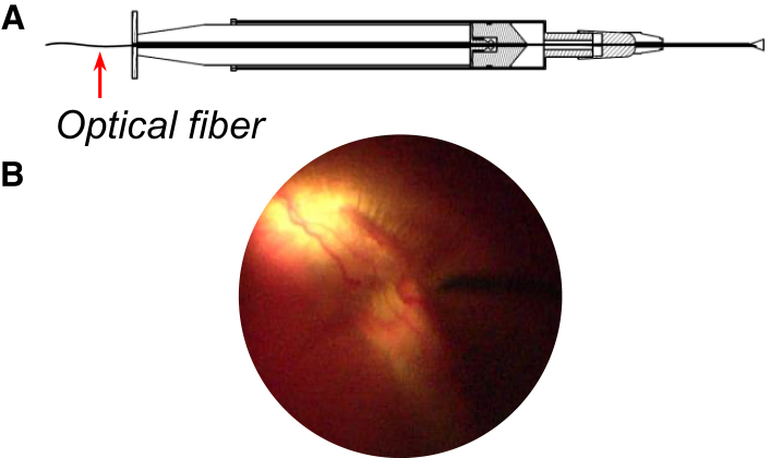
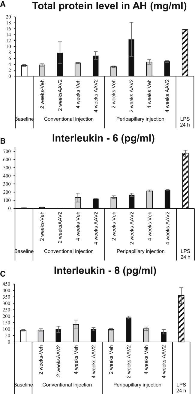
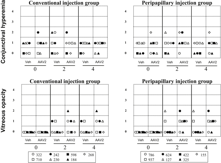
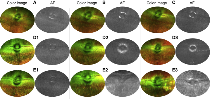
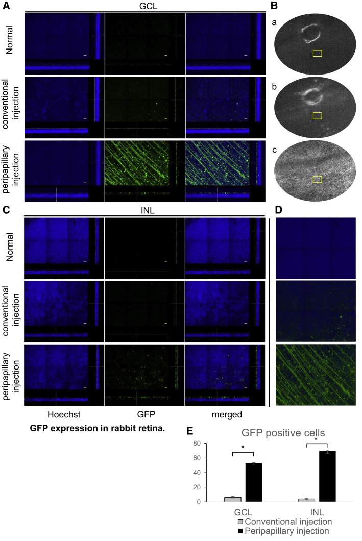
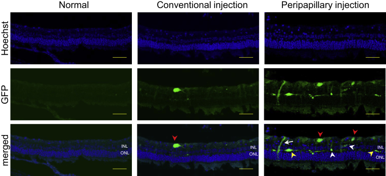
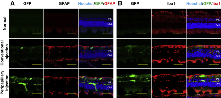
The intravitreal (IVT) injection method is a choice when targeting the inner retina for gene therapy. However, the transduction efficiency of adeno-associated virus (AAV) vectors administered by the IVT route is usually low and may be affected by several factors. To improve the transduction efficiency, we developed a novel illuminated long-needle attached injection system and injected AAV2-CMV (cytomegalovirus)-EGFP in front of the retina in rabbit eyes. Ophthalmological examinations were performed and the levels of pro-inflammatory cytokines in the aqueous humor were assessed at the baseline and 1 month, and the results were compared with those of the conventional injection method. Retinal tissues were used for immunohistochemistry. In the ophthalmological examinations, no significant inflammatory signs were detected in both groups, except for transient, mild hyperemia. In the tissues of the rabbits in the peripapillary injection group, significantly increased GFP expression was detected at the ganglion cell and the inner nuclear layers (p < 0.01). There were no differences between groups in glial activation and expressions of interleukin (IL)-6 and IL-8. These results suggest that peripapillary IVT injection in front of the retina would be safe and efficiently transduce viral vectors into the retina of large animals and is considered as a potential method for use in clinical trials.
玻璃体内(IVT)注射法是将基因疗法靶向视网膜内层时的一种选择。然而,通过IVT途径给药的腺相关病毒(AAV)载体的转导效率通常较低,并且可能受到多种因素的影响。为了提高转导效率,我们开发了一种新型的带照明长针附着注射系统,并在兔眼视网膜前方注射了AAV2-巨细胞病毒(CMV)-增强绿色荧光蛋白(EGFP)。进行了眼科检查,并在基线和1个月时评估了房水中促炎细胞因子的水平,并将结果与传统注射方法的结果进行了比较。视网膜组织用于免疫组织化学。在眼科检查中,除了短暂的轻度充血外,两组均未检测到明显的炎症迹象。在视乳头周围注射组的兔子组织中,在神经节细胞层和内核层检测到GFP表达显著增加(p<0.01)。两组在胶质细胞活化以及白细胞介素(IL)-6和IL-8的表达方面没有差异。这些结果表明,在视网膜前方进行视乳头周围IVT注射将是安全的,并且能有效地将病毒载体转导到大动物的视网膜中,被认为是一种可用于临床试验的潜在方法。