Department of Chemistry, Texas A&M University, College Station, TX 77843-3255, USA.
Metallomics. 2020 Jul 1;12(7):1094-1105. doi: 10.1039/c9mt00312f. Epub 2020 Apr 17.
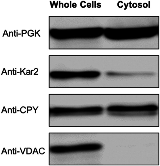
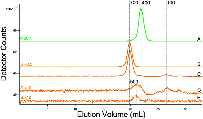
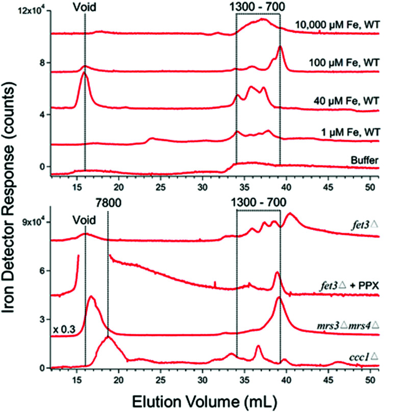
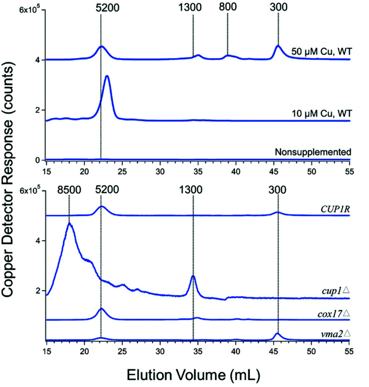
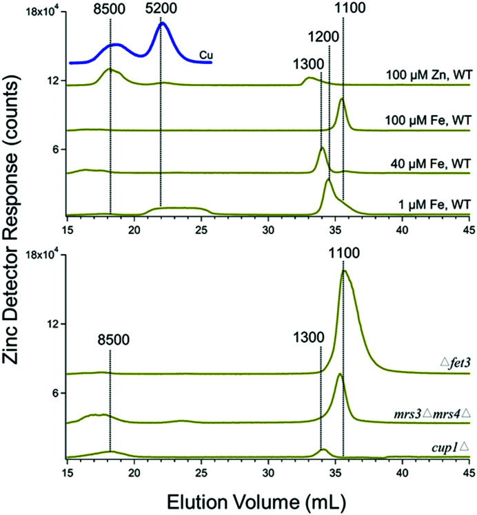
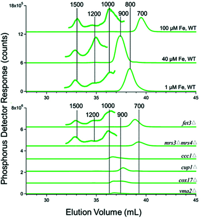
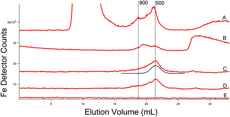
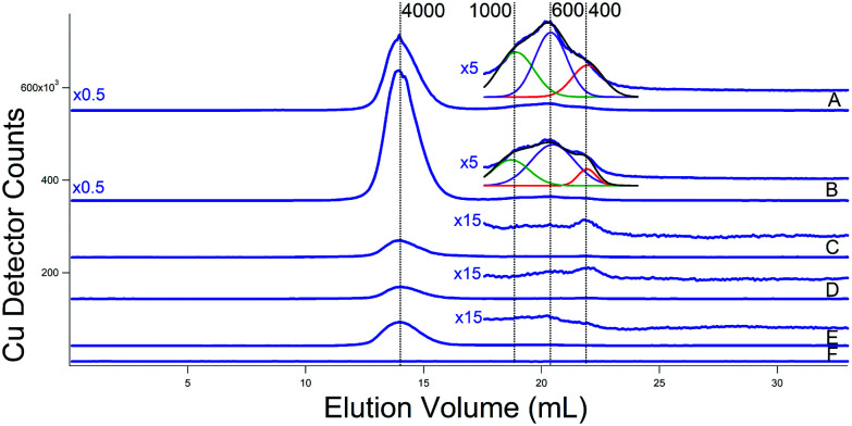
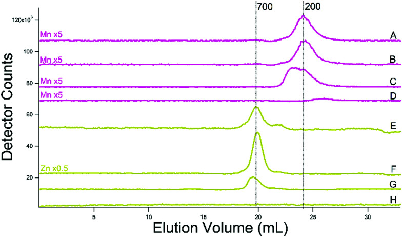
Fluorescence-based chelators are commonly used to probe labile low-molecular-mass (LMM) metal pools in the cytosol of eukaryotic cells, but such chelators destroy the complexes of interest during detection. The objective of this study was to use chromatography to directly detect such complexes. Towards this end, 47 batches of cytosol were isolated from fermenting S. cerevisiae yeast cells and passed through a 10 kDa cut-off membrane. The metal contents of the cytosol and resulting flow-through solution (FTS) were determined. FTSs were applied to a size-exclusion LC column located in an anaerobic refrigerated glove box. The LC system was coupled to an online inductively-coupled-plasma mass spectrometer (ICP-MS) for detection of individual metals. Iron-detected chromatograms of cytosolic FTSs from WT cells exhibited 2-4 major species with apparent masses between 500-1300 Da. Increasing the iron concentration in the growth medium 40-fold increased the overall intensity of these peaks. Approximately 3 LMM cytosolic copper complexes with apparent masses between 300-1300 Da were also detected; their LC intensities were weak, but these increased with increasing concentrations of copper in the growth medium. Observed higher-mass copper-detected peaks were tentatively assigned to copper-bound metallothioneins Cup1 and Crs5. FTSs from strains in which Cup1 or the Cox17 copper chaperone were deleted altered the distribution of LMM copper complexes. LMM zinc- and manganese-detected species were also present in cytosol, albeit at low concentrations. Supplementing the growth medium with zinc increased the intensity of the zinc peak assigned to Crs5 but the intensities of LMM zinc complexes were unaffected. Phosphorus-detected chromatograms were dominated by peaks at apparent masses 400-800 Da, with minor peaks at 1000-1500 Da in some batches. Sulfur chromatograms contained a low-intensity peak that comigrated with a glutathione standard; quantification suggested a GSH concentration in the cytosol of ca. 13 mM. A second LMM sulfur peak that migrated at an apparent mass of 100 Da was also evident.
基于荧光的螯合剂常用于探测真核细胞细胞质中不稳定的低分子量(LMM)金属池,但此类螯合剂在检测过程中会破坏感兴趣的复合物。本研究的目的是使用色谱法直接检测此类复合物。为此,从发酵的酿酒酵母细胞中分离了 47 批细胞质,并通过 10 kDa 截止膜。测定细胞质和所得滤液(FTS)的金属含量。FTS 应用于位于厌氧冷藏手套箱中的排阻 LC 柱。LC 系统与在线电感耦合等离子体质谱仪(ICP-MS)耦合,用于检测个别金属。WT 细胞细胞质 FTS 的铁检测色谱图显示出 2-4 种主要物质,表观质量在 500-1300 Da 之间。在生长培养基中增加铁浓度 40 倍会增加这些峰的总强度。还检测到约 3 LMM 细胞质铜复合物,表观质量在 300-1300 Da 之间;它们的 LC 强度较弱,但随着生长培养基中铜浓度的增加而增加。观察到的较高质量的铜检测峰被暂时分配给与铜结合的金属硫蛋白 Cup1 和 Crs5。Cup1 或 Cox17 铜伴侣缺失的菌株的 FTS 改变了 LMM 铜复合物的分布。LMM 锌和锰检测到的物质也存在于细胞质中,尽管浓度较低。在生长培养基中补充锌会增加分配给 Crs5 的锌峰的强度,但 LMM 锌复合物的强度不受影响。磷检测色谱图主要由表观质量为 400-800 Da 的峰主导,某些批次中在 1000-1500 Da 处有较小的峰。硫色谱图包含一个与谷胱甘肽标准品共迁移的低强度峰;定量分析表明细胞质中 GSH 浓度约为 13 mM。也明显存在另一个在表观质量为 100 Da 处迁移的 LMM 硫峰。