Center for Intelligent Signal and Imaging Research, Universiti Teknologi Petronas, Seri Iskandar, Perak, Malaysia.
Brain and Mind Research Center, Nagoya University, Nagoya, Aichi, Japan.
Hum Brain Mapp. 2020 Aug 15;41(12):3198-3211. doi: 10.1002/hbm.25008. Epub 2020 Apr 18.
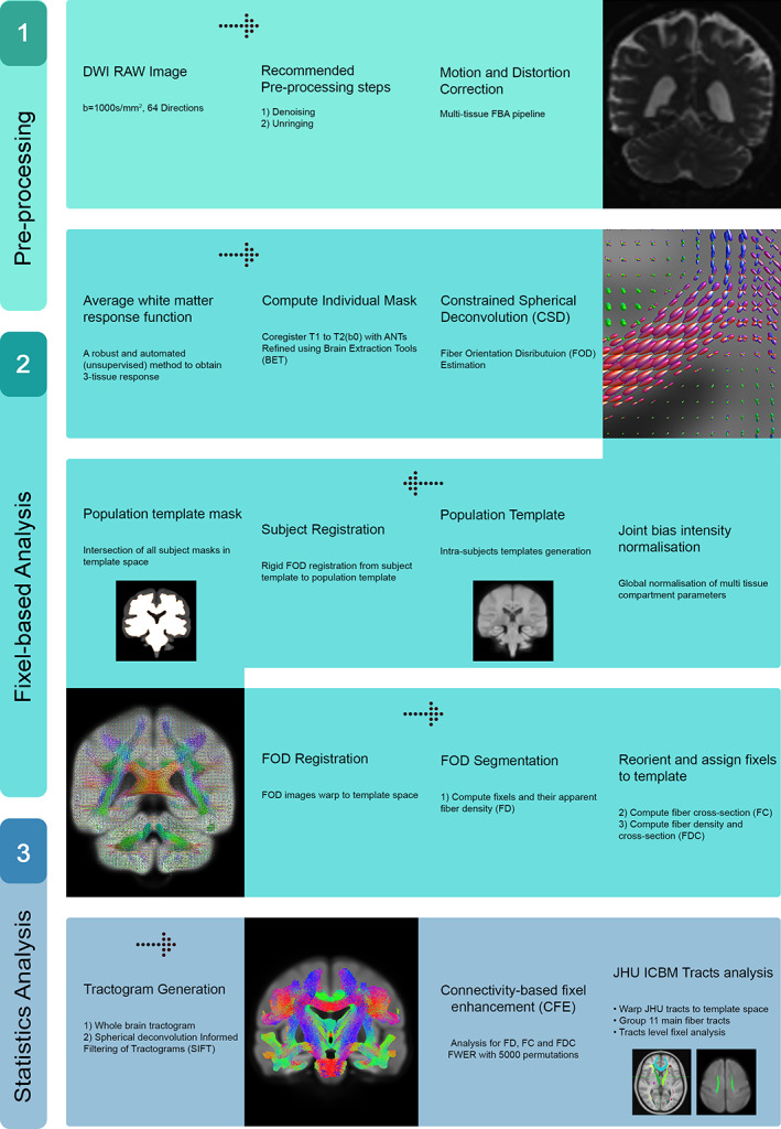
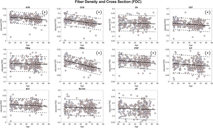
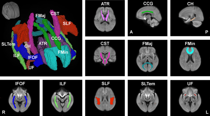
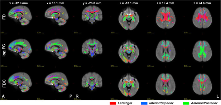
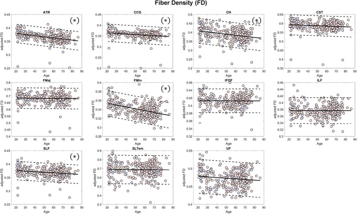
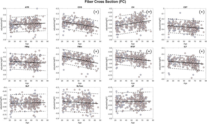
White matter (WM) fiber bundles change dynamically with age. These changes could be driven by alterations in axonal diameter, axonal density, and myelin content. In this study, we applied a novel fixel-based analysis (FBA) framework to examine these changes throughout the adult lifespan. Using diffusion-weighted images from a cohort of 293 healthy volunteers (89 males/204 females) from ages 21 to 86 years old, we performed FBA to analyze age-related changes in microscopic fiber density (FD) and macroscopic fiber morphology (fiber cross section [FC]). Our results showed significant and widespread age-related alterations in FD and FC across the whole brain. Interestingly, some fiber bundles such as the anterior thalamic radiation, corpus callosum, and superior longitudinal fasciculus only showed significant negative relationship with age in FD values, but not in FC. On the other hand, some segments of the cerebello-thalamo-cortical pathway only showed significant negative relationship with age in FC, but not in FD. Analysis at the tract-level also showed that major fiber tract groups predominantly distributed in the frontal lobe (cingulum, forceps minor) exhibited greater vulnerability to the aging process than the others. Differences in FC and the combined measure of FD and cross section values observed between sexes were mostly driven by differences in brain sizes although male participants tended to exhibit steeper negative linear relationship with age in FD as compared to female participants. Overall, these findings provide further insights into the structural changes the brain's WM undergoes due to the aging process.
脑白质(WM)纤维束随年龄而动态变化。这些变化可能是由轴突直径、轴突密度和髓鞘含量的改变驱动的。在这项研究中,我们应用了一种新的基于纤维束的分析(FBA)框架来研究整个成年期的这些变化。使用来自年龄在 21 至 86 岁的 293 名健康志愿者队列的弥散加权图像(89 名男性/204 名女性),我们进行了 FBA 以分析与年龄相关的微观纤维密度(FD)和宏观纤维形态(纤维横截面积[FC])变化。我们的结果显示,FD 和 FC 在整个大脑中都有显著且广泛的与年龄相关的改变。有趣的是,一些纤维束,如前丘脑辐射、胼胝体和上纵束,仅在 FD 值上与年龄呈显著负相关,而在 FC 上则没有。另一方面,一些小脑-丘脑-皮质通路的节段仅在 FC 上与年龄呈显著负相关,而在 FD 上则没有。在束水平的分析还表明,主要分布在额叶的纤维束组(扣带束、小内囊束)比其他纤维束组更容易受到衰老过程的影响。性别间观察到的 FC 以及 FD 和横截面积值的综合测量之间的差异主要是由脑大小的差异驱动的,尽管与女性参与者相比,男性参与者的 FD 表现出更陡峭的负线性关系。总体而言,这些发现进一步深入了解了大脑 WM 由于衰老过程而发生的结构变化。