Wu Jian, You Jieyun, Wang Xiaoyan, Wang Shijun, Huang Jiayuan, Xie Qihai, Gong Baoyong, Ding Zhiwen, Ye Yong, Wang Cong, Kang Le, Xu Ran, Li Yang, Chen Ruizhen, Sun Aijun, Yang Xiangdong, Jiang Hong, Yang Fenghua, Backx Peter H, Ge Junbo, Zou Yunzeng
Shanghai Institute of Cardiovascular Diseases, Zhongshan Hospital and Institutes of Biomedical Sciences, Fudan University, Shanghai 200032, China.
Department of Cardiovascular Medicine, Shanghai East Hospital, Tongji University School of Medicine, Shanghai 200120, China.
Ann Transl Med. 2020 Mar;8(5):219. doi: 10.21037/atm.2020.01.51.
Although aortic regurgitation (AR) is a clinically important condition that is becoming increasingly common, few relevant murine models and mechanistic studies exist for this condition. In this study, we attempted to delineate the pathological and molecular changes and address the roles of some potentially relevant molecules in an animal model of surgically induced AR.
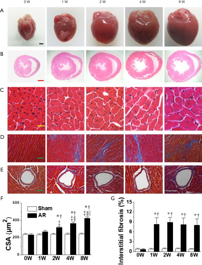
AR was induced by puncturing the aortic valve leaflets in C57BL/6J mice under echocardiographic guidance.
As early as 1 week following AR, the left ventricles (LV) displayed marked impairments in diastolic function and coronary flow reserve (CFR), as well as cardiac hypertrophy and chamber dilatation at both end-systole and end-diastole. LV free wall thickening and cardiomyocyte hypertrophy in LV were observed 2 weeks following of AR while a decline in ejection fraction was not seen until after 4 weeks. and increased over time, in conjunction with prominent Akt activation as well as slight CaMKII (Ca/calmodulin-dependent protein kinase II) activation and biphasic changes in β-arrestin-2 expression. Treatment of AR mice with Akt inhibition exacerbated the eccentric hypertrophy, while neither inhibition of CaMKII nor β-arrestin-2 overexpression influenced the response to AR.
Our structural, functional, molecular and therapeutic analyses reveal that Akt, but not CaMKII or β-arrestin-2, plays a regulatory role in the development of LV remodeling after AR in Mice. These results may shed important light on therapeutic targets for volume overloaded cardiomyopathy.
尽管主动脉瓣反流(AR)是一种临床上重要且日益常见的病症,但针对该病症的相关小鼠模型和机制研究却很少。在本研究中,我们试图在手术诱导的AR动物模型中描绘病理和分子变化,并探讨一些潜在相关分子的作用。
在超声心动图引导下,通过穿刺C57BL/6J小鼠的主动脉瓣叶来诱导AR。
早在AR发生后1周,左心室(LV)的舒张功能和冠状动脉血流储备(CFR)就出现明显受损,同时在收缩末期和舒张末期均出现心脏肥大和心室扩张。AR发生2周后观察到LV游离壁增厚和LV心肌细胞肥大,而射血分数直到4周后才下降。 并随时间增加,同时伴有显著的Akt激活以及轻微的CaMKII(钙/钙调蛋白依赖性蛋白激酶II)激活和β-抑制蛋白2表达的双相变化。用Akt抑制剂治疗AR小鼠会加剧离心性肥大,而抑制CaMKII或过表达β-抑制蛋白2均不影响对AR的反应。
我们的结构、功能、分子和治疗分析表明,在小鼠AR后LV重塑的发展中起调节作用的是Akt,而非CaMKII或β-抑制蛋白2。这些结果可能为容量超负荷性心肌病的治疗靶点提供重要线索。