Craciun Constantin
Babes-Bolyai University, Cluj-Napoca, Romania.
Discoveries (Craiova). 2014 Aug 5;2(3):e23. doi: 10.15190/d.2014.15.
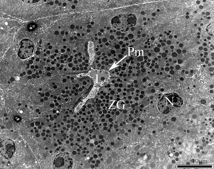
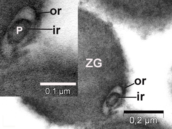
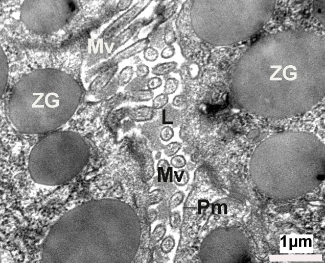
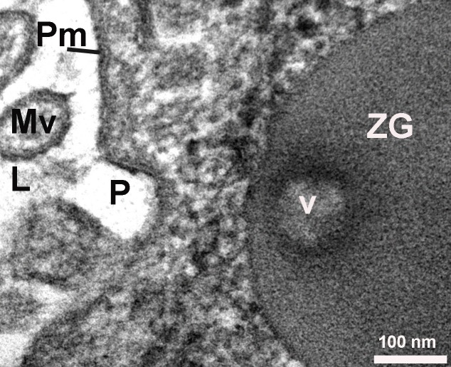
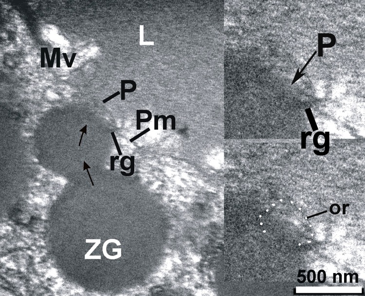
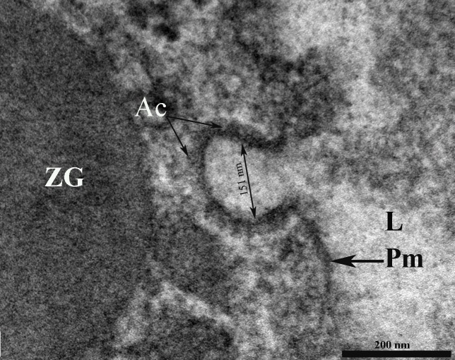
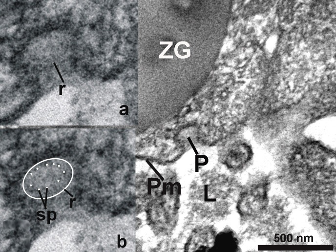
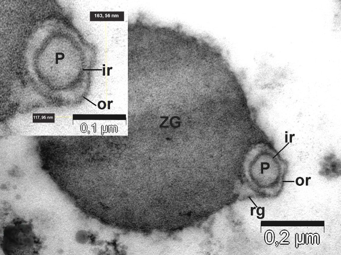
A major question in cell biology that accumulation of partially empty vesicles in cells following secretion is seen, while it is believed that secretion occurs via the complete merger of secretory vesicles with the cell plasma membrane. This important question was solved nearly two decades ago, with the discovery of the Porosome. Porosomes are cup-shaped lipoprotein structures found at the plasma membrane of all cells. Secretory vesicles dock and transiently fuse at the porosome base to form a continuous channel or fusion pore to release the pressurized vesicle contents to the outside. In a decade-long study by our group, we carefully examined using electron microscopy, the detailed structure of the porosome complex in acinar cells of the exocrine pancreas. Besides conformation of earlier findings, our study provides in much greater detail, the in situ morphology of the porosome complex in the exocrine pancreas. The discovery of the detailed morphology of the exocrine pancreas porosome complex in my laboratory is one of the major highlights of my academic career spanning nearly 50 years. Results from our EM studies, reveal for the first time the presence of tethers or cables, which are likely t-SNAREs, present at the porosome base. These EM studies further demonstrate for the first time the docking of a single secretory vesicle or zymogen granule at the base of more than one porosome complex. Detailed spoke-like elements lining the porosome cup were also observed for the first time in these studies, greatly advancing our understanding of the molecular architecture and physiology of the porosome in the exocrine pancreas.
细胞生物学中的一个主要问题是,在分泌后细胞内会出现部分排空的囊泡积累现象,而人们认为分泌是通过分泌囊泡与细胞质膜的完全融合来进行的。这个重要问题在近二十年前随着孔体的发现而得到解决。孔体是在所有细胞的质膜上发现的杯状脂蛋白结构。分泌囊泡在孔体底部对接并短暂融合,形成一个连续的通道或融合孔,将加压的囊泡内容物释放到细胞外。在我们小组长达十年的研究中,我们使用电子显微镜仔细检查了外分泌胰腺腺泡细胞中孔体复合物的详细结构。除了证实早期的发现外,我们的研究更详细地提供了外分泌胰腺中孔体复合物的原位形态。在我近50年的学术生涯中,我实验室对外分泌胰腺孔体复合物详细形态的发现是主要亮点之一。我们的电子显微镜研究结果首次揭示了在孔体底部存在可能是t-SNARE的系链或索状物。这些电子显微镜研究还首次进一步证明了单个分泌囊泡或酶原颗粒在不止一个孔体复合物底部的对接。在这些研究中还首次观察到了排列在孔体杯周围的详细的辐状元件,极大地推进了我们对外分泌胰腺中孔体分子结构和生理学的理解。