Laboratory of Structural Biology Research, National Institute of Arthritis and Musculoskeletal and Skin Diseases, National Institutes of Health, Bethesda, Maryland, United States of America.
Protein Expression Laboratory, National Institute of Arthritis and Musculoskeletal and Skin Diseases, National Institutes of Health, Bethesda, Maryland, United States of America.
PLoS Comput Biol. 2020 Apr 20;16(4):e1007782. doi: 10.1371/journal.pcbi.1007782. eCollection 2020 Apr.
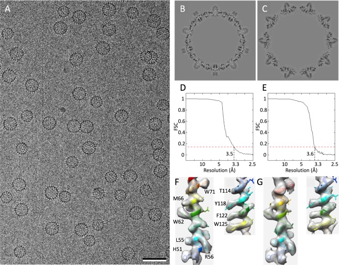
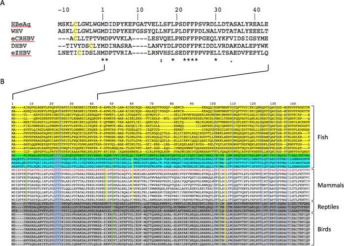
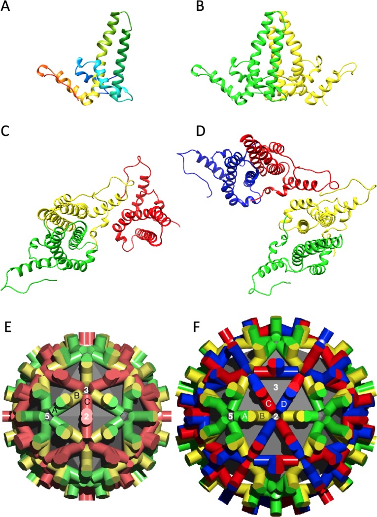
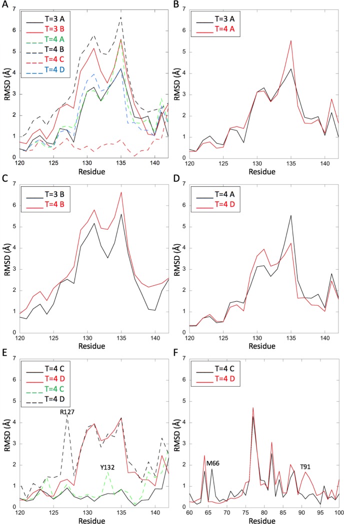
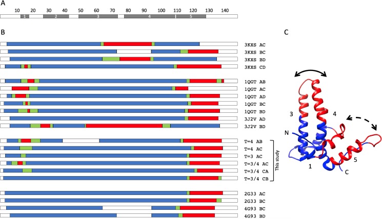
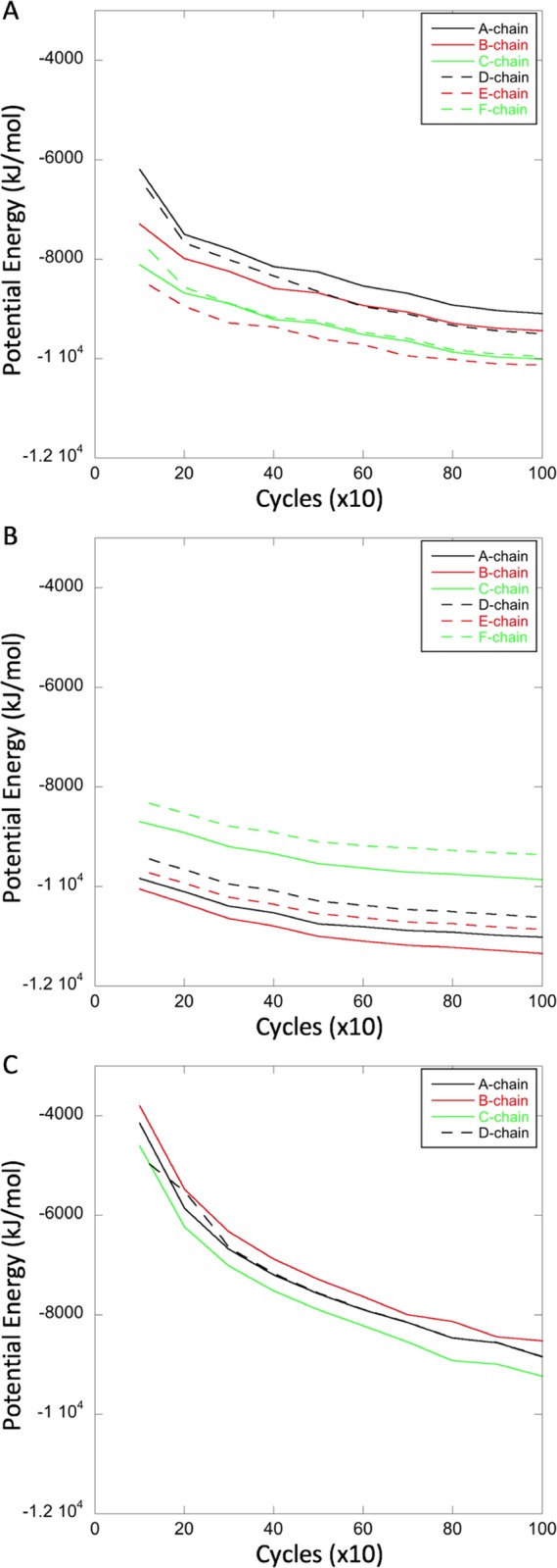
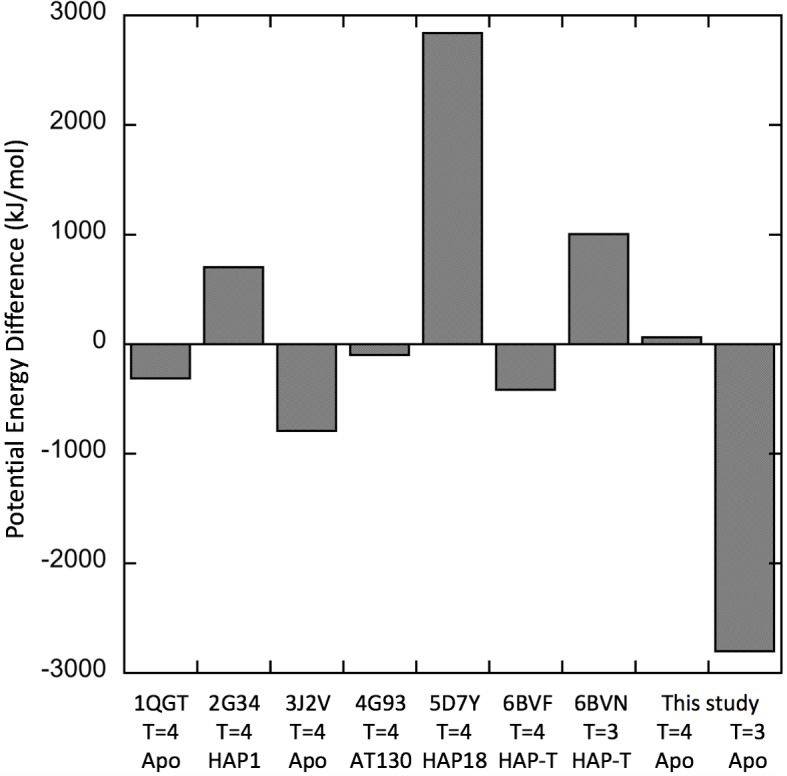
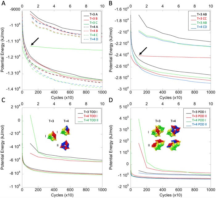
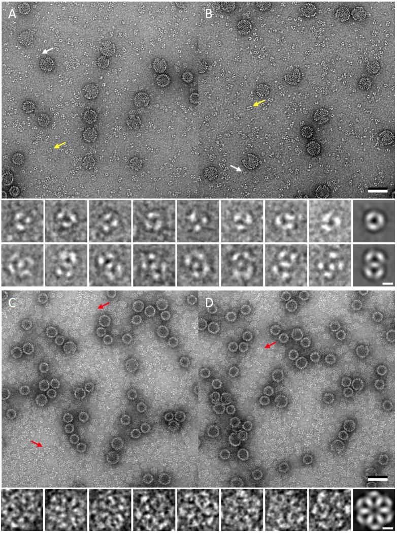
Hepatitis B virus (HBV) is a leading cause of liver disease. The capsid is an essential component of the virion and it is therefore of interest how it assembles and disassembles. The capsid protein is unusual both for its rare fold and that it polymerizes according to two different icosahedral symmetries, causing the polypeptide chain to exist in seven quasi-equivalent environments: A, B, and C in AB and CC dimers in T = 3 capsids, and A, B, C, and D in AB and CD dimers in T = 4 capsids. We have compared the two capsids by cryo-EM at 3.5 Å resolution. To ensure a valid comparison, the two capsids were prepared and imaged under identical conditions. We find that the chains have different conformations and potential energies, with the T = 3 C chain having the lowest. Three of the four quasi-equivalent dimers are asymmetric with respect to conformation and potential energy; however, the T = 3 CC dimer is symmetrical and has the lowest potential energy although its intra-dimer interface has the least free energy of formation. Of all the inter-dimer interfaces, the CB interface has the least area and free energy, in both capsids. From the calculated energies of higher-order groupings of dimers discernible in the lattices we predict early assembly intermediates, and indeed we observe such structures by negative stain EM of in vitro assembly reactions. By sequence analysis and computational alanine scanning we identify key residues and motifs involved in capsid assembly. Our results explain several previously reported observations on capsid assembly, disassembly, and dimorphism.
乙型肝炎病毒 (HBV) 是肝脏疾病的主要病因。衣壳是病毒粒子的重要组成部分,因此研究其组装和解组装方式很有意义。衣壳蛋白的折叠方式非常罕见,而且它根据两种不同的二十面体对称性聚合,导致多肽链存在于七个准等效环境中:AB 二聚体中的 A、B 和 C 以及 T = 3 衣壳中的 CC 二聚体中的 A、B 和 C;AB 和 CD 二聚体中的 A、B、C 和 D 在 T = 4 衣壳中。我们通过 cryo-EM 在 3.5 Å 分辨率下比较了这两种衣壳。为了确保进行有效的比较,我们在相同条件下制备和成像了这两种衣壳。我们发现,这些链具有不同的构象和势能,T = 3 C 链的势能最低。四个准等效二聚体中的三个在构象和势能方面是不对称的;然而,T = 3 CC 二聚体是对称的,并且具有最低的势能,尽管其内部二聚体界面的形成自由能最小。在所有的二聚体界面中,CB 界面在两种衣壳中的面积和自由能最小。从晶格中可辨别的更高阶二聚体的计算能量中,我们预测了早期的组装中间体,并且实际上我们通过体外组装反应的负染电镜观察到了这些结构。通过序列分析和计算丙氨酸扫描,我们确定了参与衣壳组装的关键残基和基序。我们的结果解释了以前关于衣壳组装、解组装和二态性的一些报告观察结果。