Department of Cell Physiology, The Jikei University School of Medicine, Tokyo, Japan.
Department of Life Science and Medical Bioscience, Waseda University, Tokyo, Japan.
PLoS One. 2020 Apr 21;15(4):e0231905. doi: 10.1371/journal.pone.0231905. eCollection 2020.
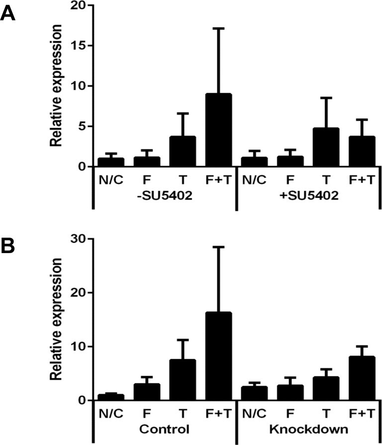
Myocardial fibrosis is often associated with cardiac hypertrophy; indeed, fibrosis is one of the most critical factors affecting prognosis. We aimed to identify the molecules involved in promoting fibrosis under hypertrophic stimuli. We previously established a rat model of cardiac hypertrophy by pulmonary artery banding, in which approximately half of the animals developed fibrosis in the right ventricle. Here, we first comprehensively analyzed mRNA expression in the right ventricle with or without fibrosis in pulmonary artery banding model rats by DNA microarray analysis (GSE141650 at NCBI GEO). The expression levels of 19 genes were up-regulated more than 1.5-fold in fibrotic hearts compared with non-fibrotic hearts. Among them, fibrosis growth factor (FGF) 23 showed one of the biggest increases in expression. Real-time PCR analysis also revealed that, among the FGF receptor (FGFR) family, FGFR1 was highly expressed in fibrotic hearts. We then found that FGF23 was expressed predominantly in cardiomyocytes, while FGFR1 was predominantly expressed in fibroblasts in the rat ventricle. Next, we added FGF23 and transforming growth factor (TGF)-β1 (10-50 ng/mL of each) to isolated fibroblasts from normal adult rat ventricles and cultured them for three days. While FGF23 itself did not directly affect the expression levels of any fibrosis-related mRNAs, FGF23 enhanced the effect of TGF-β1 on increasing the expression levels of α-smooth muscle actin (α-SMA) mRNA. This increase in xx-SMA mRNA levels due to the combination of TGF-β1 and FGF23 was attenuated by the inhibition of FGFR1 or the knockdown of FGFR1 in fibroblasts. Thus, FGF23 synergistically promoted the activation of fibroblasts with TGF-β1, transforming fibroblasts into myofibroblasts via FGFR1. Thus, we identified FGF23 as a paracrine factor secreted from cardiomyocytes to promote cardiac fibrosis under conditions in which TGF-β1 is activated. FGF23 could be a possible target to prevent fibrosis following myocardial hypertrophy.
心肌纤维化通常与心脏肥大有关;事实上,纤维化是影响预后的最重要因素之一。我们旨在确定在肥大刺激下促进纤维化的相关分子。我们之前通过肺动脉结扎建立了大鼠心脏肥大模型,其中大约一半的动物在右心室发生纤维化。在这里,我们首先通过 DNA 微阵列分析(NCBI GEO 中的 GSE141650)全面分析了肺动脉结扎模型大鼠的右心室纤维化或无纤维化组织的 mRNA 表达。与无纤维化心脏相比,纤维化心脏中 19 个基因的表达水平上调了 1.5 倍以上。其中,纤维化生长因子(FGF)23 的表达增加最为显著。实时 PCR 分析还显示,在 FGF 受体(FGFR)家族中,FGFR1 在纤维化心脏中高表达。我们发现 FGF23 主要在心肌细胞中表达,而 FGFR1 主要在大鼠心室成纤维细胞中表达。接下来,我们向正常成年大鼠心室分离的成纤维细胞中添加 FGF23 和转化生长因子(TGF)-β1(每种 10-50ng/ml),并培养它们 3 天。虽然 FGF23 本身不会直接影响任何纤维化相关 mRNAs 的表达水平,但 FGF23 增强了 TGF-β1 增加α-平滑肌肌动蛋白(α-SMA)mRNA 表达水平的作用。由于 TGF-β1 和 FGF23 的组合,这种 α-SMA mRNA 水平的增加被 FGFR1 抑制或成纤维细胞中 FGFR1 的敲低所减弱。因此,FGF23 与 TGF-β1 协同促进成纤维细胞的激活,通过 FGFR1 将成纤维细胞转化为肌成纤维细胞。因此,我们确定 FGF23 是一种旁分泌因子,可在 TGF-β1 激活的情况下从心肌细胞分泌,促进心脏纤维化。FGF23 可能是预防心肌肥大后纤维化的一个可能靶点。