Molecular Oncology and Genetics Department, Innovative Medical Forum, The F. Lukaszczyk Oncology Center, 85-796 Bydgoszcz, Poland.
Department of Thoracic Surgery and Tumors, Ludwik Rydygier Collegium Medicum in Bydgoszcz, Nicolaus Copernicus University, 85-067 Torun, Poland.
Molecules. 2020 Apr 17;25(8):1864. doi: 10.3390/molecules25081864.
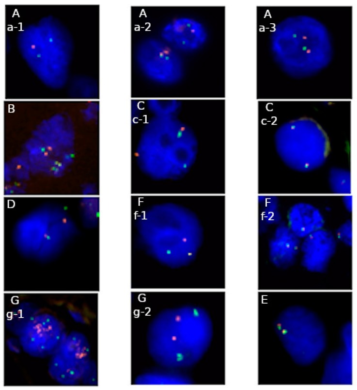
Fluorescence in situ hybridization (FISH) is a standard technique used in routine diagnostics of genetic aberrations. Thanks to simple FISH procedure is possible to recognize tumor-specific abnormality. Its applications are limited to designed probe type. Gene rearrangements e.g., , reflecting numerous translocational partners, deletions of critical regions e.g., 1p and 19q, gene fusions e.g., , genomic imbalances e.g., 6p, 6q, 11q and amplifications e.g., are targets in personalized oncology. Confirmation of genetic marker is frequently a direct indication to start specific, targeted treatment. In other cases, detected aberration helps pathologists to better distinguish soft tissue sarcomas, or to state a final diagnosis. Our main goal is to show that applying FISH to formalin-fixed paraffin-embedded tissue sample (FFPE) enables assessing genomic status in the population of cells deriving from a primary tumor or metastasis. Although many more sophisticated techniques are available, like Real-Time PCR or new generation sequencing, FISH remains a commonly used method in many genetic laboratories.
荧光原位杂交(FISH)是一种用于遗传异常常规诊断的标准技术。由于 FISH 方法简单,因此可以识别肿瘤特异性异常。其应用仅限于设计的探针类型。基因重排,例如,反映许多易位伙伴,关键区域缺失,例如 1p 和 19q,基因融合,例如,基因组不平衡,例如 6p、6q、11q 和扩增,例如 ,是个性化肿瘤学的靶点。遗传标志物的确认通常是开始特定靶向治疗的直接指征。在其他情况下,检测到的异常有助于病理学家更好地区分软组织肉瘤,或作出最终诊断。我们的主要目标是表明,将 FISH 应用于福尔马林固定石蜡包埋组织样本(FFPE)能够评估源自原发性肿瘤或转移灶的细胞群体中的基因组状态。尽管有许多更复杂的技术,如实时 PCR 或新一代测序,但 FISH 仍然是许多遗传实验室常用的方法。