Deken Marion M, Kijanka Marta M, Beltrán Hernández Irati, Slooter Maxime D, de Bruijn Henriette S, van Diest Paul J, van Bergen En Henegouwen Paul M P, Lowik Clemens W G M, Robinson Dominic J, Vahrmeijer Alexander L, Oliveira Sabrina
Dept. of Surgery, Leiden University Medical Center, Leiden, the Netherlands.
Division of Cell Biology, Neurobiology and Biophysics, Dept. of Biology, Faculty of Science, Utrecht University, Utrecht, the Netherlands.
J Control Release. 2020 Jul 10;323:269-281. doi: 10.1016/j.jconrel.2020.04.030. Epub 2020 Apr 21.
A substantial number of breast cancer patients with an overexpression of the human epidermal growth factor receptor 2 (HER2) have residual disease after neoadjuvant therapy or become resistant to trastuzumab. Photodynamic therapy (PDT) using nanobodies targeted to HER2 is a promising treatment option for these patients. Here we investigate the in vitro and in vivo antitumor efficacy of HER2-targeted nanobody-photosensitizer (PS) conjugate PDT.
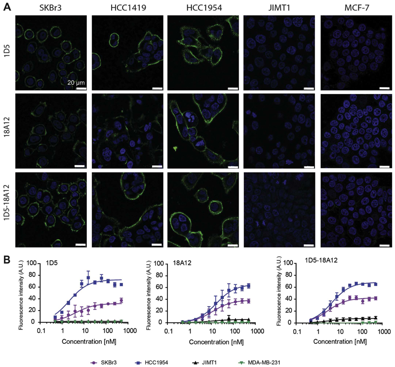
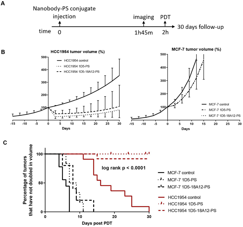
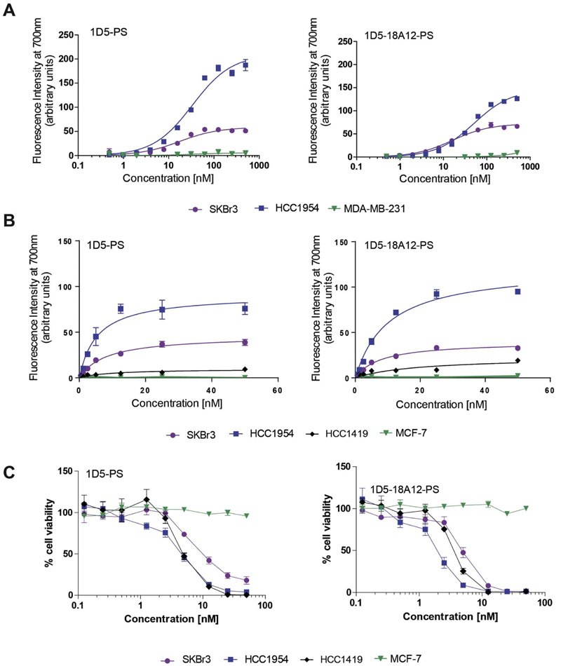
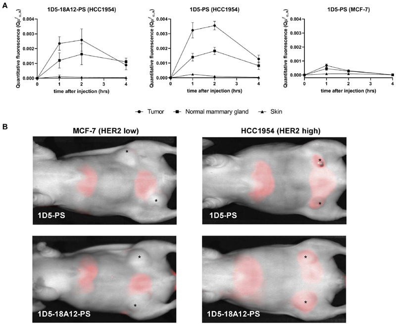
Nanobodies targeting HER2 were obtained from phage display selections. Monovalent nanobodies were engineered into a biparatopic construct. The specificity of selected nanobodies was tested in immunofluorescence assays and their affinity was evaluated in binding studies, both performed in a panel of breast cancer cells varying in HER2 expression levels. The selected HER2-targeted nanobodies 1D5 and 1D5-18A12 were conjugated to the photosensitizer IRDye700DX and tested in in vitro PDT assays. Mice bearing orthotopic HCC1954 trastuzumab-resistant tumors with high HER2 expression or MCF-7 tumors with low HER2 expression were intravenously injected with nanobody-PS conjugates. Quantitative fluorescence spectroscopy was performed for the determination of the local pharmacokinetics of the fluorescence conjugates. After nanobody-PS administration, tumors were illuminated to a fluence of 100 J∙cm, with a fluence rate of 50 mW∙cm, and thereafter tumor growth was measured with a follow-up until 30 days.
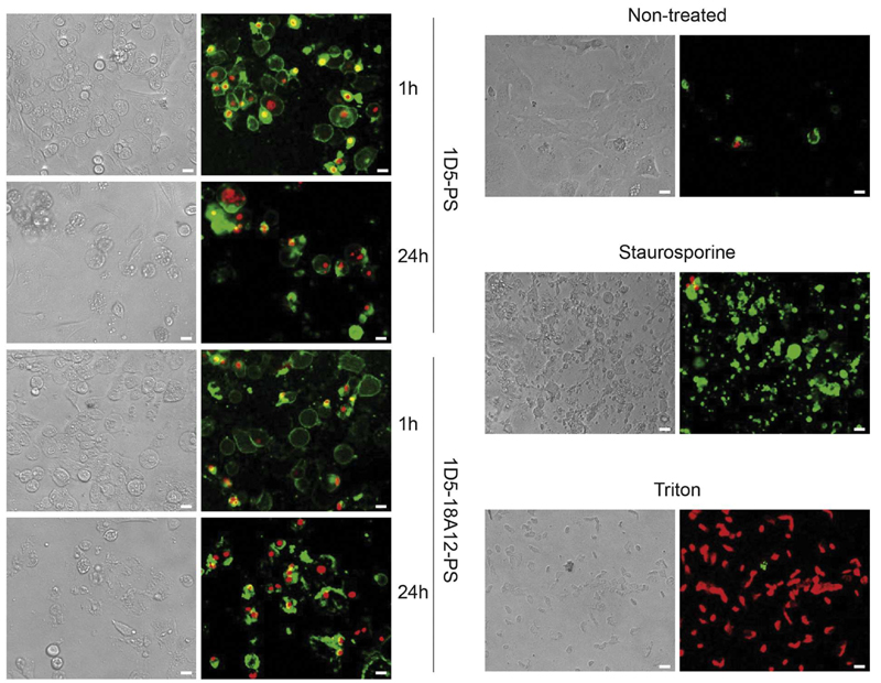
The selected nanobodies remained functional after conjugation to the PS, binding specifically and with high affinity to HER2-positive cells. Both nanobody-PS conjugates potently and selectively induced cell death of HER2 overexpressing cells, either sensitive or resistant to trastuzumab, with low nanomolar LD values. In vivo, quantitative fluorescence spectroscopy showed specific accumulation of nanobody-PS conjugates in HCC1954 tumors and indicated 2 h post injection as the most suitable time point to apply light. Nanobody-targeted PDT with 1D5-PS and 1D5-18A12-PS induced significant tumor regression of trastuzumab-resistant high HER2 expressing tumors, whereas in low HER2 expressing tumors only a slight growth delay was observed.
Nanobody-PS conjugates accumulated selectively in vivo and their fluorescence could be detected through optical imaging. Upon illumination, they selectively induced significant tumor regression of HER2 overexpressing tumors with a single treatment session. Nanobody-targeted PDT is therefore suggested as a new additional treatment for HER2-positive breast cancer, particularly of interest for trastuzumab-resistant HER2-positive breast cancer. Further studies are now needed to assess the value of this approach in clinical practice.
大量人表皮生长因子受体2(HER2)过表达的乳腺癌患者在新辅助治疗后仍有残留疾病或对曲妥珠单抗产生耐药性。使用靶向HER2的纳米抗体的光动力疗法(PDT)是这些患者一种有前景的治疗选择。在此,我们研究靶向HER2的纳米抗体-光敏剂(PS)偶联物PDT的体外和体内抗肿瘤疗效。
从噬菌体展示筛选中获得靶向HER2的纳米抗体。将单价纳米抗体改造成双特异性结构。在免疫荧光试验中测试所选纳米抗体的特异性,并在结合研究中评估其亲和力,这两项研究均在一组HER2表达水平不同的乳腺癌细胞中进行。将所选的靶向HER2的纳米抗体1D5和1D5-18A12与光敏剂IRDye700DX偶联,并在体外PDT试验中进行测试。对携带HER2高表达的原位HCC1954曲妥珠单抗耐药肿瘤或HER2低表达的MCF-7肿瘤的小鼠静脉注射纳米抗体-PS偶联物。进行定量荧光光谱分析以确定荧光偶联物的局部药代动力学。在给予纳米抗体-PS后,以50 mW∙cm的光通量率将肿瘤照射至100 J∙cm的光通量,然后测量肿瘤生长情况并随访30天。
所选纳米抗体与PS偶联后仍保持功能,能特异性且高亲和力地结合HER2阳性细胞。两种纳米抗体-PS偶联物均能有效且选择性地诱导HER2过表达细胞(无论对曲妥珠单抗敏感还是耐药)的细胞死亡,其半数致死剂量值低至纳摩尔级别。在体内,定量荧光光谱显示纳米抗体-PS偶联物在HCC1954肿瘤中特异性蓄积,并表明注射后2小时是施加光照的最合适时间点。用1D5-PS和1D5-18A12-PS进行的纳米抗体靶向PDT可诱导HER2高表达的曲妥珠单抗耐药肿瘤显著消退,而在HER2低表达肿瘤中仅观察到轻微生长延迟。
纳米抗体-PS偶联物在体内选择性蓄积,其荧光可通过光学成像检测到。光照后,它们通过单次治疗即可选择性地诱导HER2过表达肿瘤显著消退。因此,纳米抗体靶向PDT被认为是HER2阳性乳腺癌的一种新的附加治疗方法,对于曲妥珠单抗耐药的HER2阳性乳腺癌尤其有意义。现在需要进一步研究来评估这种方法在临床实践中的价值。